Review
. 2023 Aug 10;22(1):130.
doi: 10.1186/s12943-023-01831-w.

Multifaceted role of redox pattern in the tumor immune microenvironment regarding autophagy and apoptosis

Affiliations
Review

Multifaceted role of redox pattern in the tumor immune microenvironment regarding autophagy and apoptosis

Yuqing Ren et al. Mol Cancer. .

Abstract

The reversible oxidation-reduction homeostasis mechanism functions as a specific signal transduction system, eliciting related physiological responses. Disruptions to redox homeostasis can have negative consequences, including the potential for cancer development and progression, which are closely linked to a series of redox processes, such as adjustment of reactive oxygen species (ROS) levels and species, changes in antioxidant capacity, and differential effects of ROS on downstream cell fate and immune capacity. The tumor microenvironment (TME) exhibits a complex interplay between immunity and regulatory cell death, especially autophagy and apoptosis, which is crucially regulated by ROS. The present study aims to investigate the mechanism by which multi-source ROS affects apoptosis, autophagy, and the anti-tumor immune response in the TME and the mutual crosstalk between these three processes. Given the intricate role of ROS in controlling cell fate and immunity, we will further examine the relationship between traditional cancer therapy and ROS. It is worth noting that we will discuss some potential ROS-related treatment options for further future studies.

Keywords: Apoptosis; Autophagy; Cancer therapy; Immunity; Redox and ROS.


Conflict of interest statement

The authors declare that they have no competing interests.

Figures
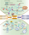
Fig. 1
Fig. 1
Mechanism and effect of autophagy and apoptosis. Autophagy and apoptosis are two vital mechanisms for maintaining cellular homeostasis under stress conditions. Autophagy is an evolutionarily conserved cellular degradation process that is activated in response to cellular stress signals. The ULK1 complex mediates autophagy initiation, and phagocytic vacuole formation involves the class III phosphoinositide 3-kinase (PI3K) complex, comprising PI3K, ATG14L, Beclin 1, VPS34, and VPS15. The ATG5/ATG12/ATG16 complex and LC3II are subsequently involved, and during autophagosome formation, p62 binds to LC3II while the phagophore expands, encapsulating intracellular material to form autophagosomes. Lysosomes fuse with autophagosomes, providing hydrolytic enzymes for the degradation of phagocytosed material. Two signaling pathways for apoptosis exist: intrinsic and extrinsic. Various intracellular microenvironment perturbations such as DNA damage, growth factor deprivation, and oxidative stress activate the intrinsic apoptotic pathway. Mitochondrial outer membrane permeabilization (MOMP) is a critical step in apoptosis, leading to the release of intermembrane proteins such as cytochrome c. Proteins of the BCL family promote or inhibit this process. Cytochrome c binds to Apaf-1 to form apoptotic bodies, promoting caspase activation. In contrast, extrinsic apoptosis is triggered by the binding of death ligands (e.g., FasL, TNF-α) to death receptors (e.g., Fas, TNF-R), resulting in the assembly of death-inducing signaling complexes (DISC) and the activation of downstream effector caspases (e.g., caspases 3, 6, 7, 8, 10). The two play different roles at different stages
Fig. 2
Fig. 2
ROS Generation and Signaling Mechanisms in Tumor Microenvironment Dynamics.Mitochondrial ROS are predominantly generated through the mitochondrial electrical transport chain (ETC), primarily complex I (Com I) and Com III. Other mitochondrial enzymes, including NADPH oxidase (NOX), can also directly catalyze ROS generation via enzymatic reactions. NOX proteins require various regulatory subunits, includingp40phox,p47phox and p67phox, to exert catalytic effects. These subunits have multiple components in the tumor microenvironment, with differing ROS production and action pathways. In APC-deficient colorectal cancer cells, ROS production and NF-κB activation in the NOX pathway, triggered by RAC1, promote WNT-driven intestinal stem cell proliferation and cancer development. The mitochondrial ROS pathway can regulate tumor cell proliferation and growth by activating kinases such as JNK and p38 MAPK, as well as ATM in the DNA damage pathway. Cancer-associated fibroblasts (CAFs) in the tumor microenvironment promote tumor growth and progression and produce ROS through downregulation of mitochondrial electron transport chain, NOX, and antioxidant mechanisms. Oxidative stress can convert fibroblasts into myofibroblasts. ROS mediate TGF-β signaling through various pathways, including redox-dependent accumulation of hypoxia-inducible factor (HIF), stimulating the SDF-1/CXCR4 signaling pathway and activating RhoA-GTPase. Alternatively, ROS can independently stimulate SDF-1, which then causes myofibroblast characteristics in immune cells of TME. ROS production is mainly by one of the two modalities listed above, depending on the cell type, and is regulated by different stimulating factors and signaling pathways. See the figure for a detailed list of these factors and pathways
Fig. 3
Fig. 3
Role of ROS in innate immunity. ROS have a dual role in innate immunity and can function as either a potential therapeutic target against cancer by inhibiting tumor cell proliferation and promoting innate immune cells or as an obstacle to treatment by promoting tumor cell proliferation and inhibiting innate immune cells. a Promotion. ROS contribute to macrophages becoming M1 phenotype and mediating the phagocytic activity of M1 macrophages by releasing factors such as IL-1, IL-12, and TNF-α. Neutrophils play a crucial role in killing tumor cells and preventing tumor spread and metastasis. ROS generation induces excessive calcium influx in tumor cells through TRPM2 channels, inhibiting tumor cell growth and metastasis and driving the cytotoxic effect of neutrophils. ROS production is an early event after NK cells recognize cancer cells and induce the production of cytotoxic essential substances such as perforin and granzyme. ROS generation in DC cells regulates their phagocytic activity and maintains alkalinization. When tumor cells enter DCs, the activation of STING by tumor cell DNA promotes the production of IFN1 and other defense cytokines. b Suppression. ROS promote macrophage transformation to M2 type and secrete inhibitory cytokines such as IL-10 and TGF-β, which have pro-angiogenic and immunosuppressive functions and promote tumorigenesis. ROS also promote the generation of neutrophil extracellular traps (NETs), which can have both tumor-promoting and metastasis-promoting effects. High ROS levels in the TME may also be detrimental to NK cell survival, and L-kynurenine as well as lactate produced by IDO can lead to NK cell apoptosis through ROS pathways in NK cells. Additionally, excess ROS can oxidize lipids and form lipid bodies (LB) of electrophilic oxidized truncated (ox-tr) lipids, inhibiting antigen presentation by DCs to T cells and weakening immune responses
Fig. 4
Fig. 4
Role of ROS in adaptive immunity. Adaptive immunity in tumors involves the capture, processing, and presentation of antigens by DCs, leading to the activation of effector T cells against specific antigens. This process is linked to ROS in several ways. The release of cancer antigens is regulated by the type of cell death, with autophagy or apoptotic cell death determining the release of cancer antigens. The amount of ROS present during antigen presentation affects its efficacy, with inhibition of ROS significantly reducing antigen uptake by DCs. However, modest ROS levels are required for T cell activation and differentiation. NOX2-derived ROS are involved in CD3/CD28 stimulation-mediated CD8 + T cell activation, and TCR activation promotes T cell activation by inducing ROS production and regulating IL2 and IL4 expression. Activated T cells express chemokines in response to ROS, which facilitate their migration to the tumor site, where they can induce apoptosis by expressing death ligands such as FasL and TRAIL. ROS are also involved in IL-2-dependent IL-2 production, and subsequent TNF, IFN-γ, perforin, and granzyme B production, as TCR signaling is sensitive to ROS

Similar articles

Cited by

References

    1. Sies H, Jones DP. Reactive oxygen species (ROS) as pleiotropic physiological signalling agents. Nat Rev Mol Cell Biol. 2020;21:363–83. doi: 10.1038/s41580-020-0230-3. - DOI - PubMed
    1. D’Autréaux B, Toledano MB. ROS as signalling molecules: mechanisms that generate specificity in ROS homeostasis. Nat Rev Mol Cell Biol. 2007;8:813–24. doi: 10.1038/nrm2256. - DOI - PubMed
    1. van der Pol A, van Gilst WH, Voors AA, van der Meer P. Treating oxidative stress in heart failure: past, present and future. Eur J Heart Fail. 2019;21:425–35. doi: 10.1002/ejhf.1320. - DOI - PMC - PubMed
    1. Cheung EC, Vousden KH. The role of ROS in tumour development and progression. Nat Rev Cancer. 2022;22:280–97. doi: 10.1038/s41568-021-00435-0. - DOI - PubMed
    1. Belli C, Trapani D, Viale G, D’Amico P, Duso BA, Della Vigna P, Orsi F, Curigliano G. Targeting the microenvironment in solid tumors. Cancer Treat Rev. 2018;65:22–32. doi: 10.1016/j.ctrv.2018.02.004. - DOI - PubMed

Publication types

Substances