. 2023 Aug 10;20(1):13.
doi: 10.1186/s12977-023-00628-5.

Transmitted/founder SHIV.D replicates in the brain, causes neuropathogenesis, and persists on combination antiretroviral therapy in rhesus macaques

Affiliations

Transmitted/founder SHIV.D replicates in the brain, causes neuropathogenesis, and persists on combination antiretroviral therapy in rhesus macaques

Rachel M Podgorski et al. Retrovirology. .

Abstract

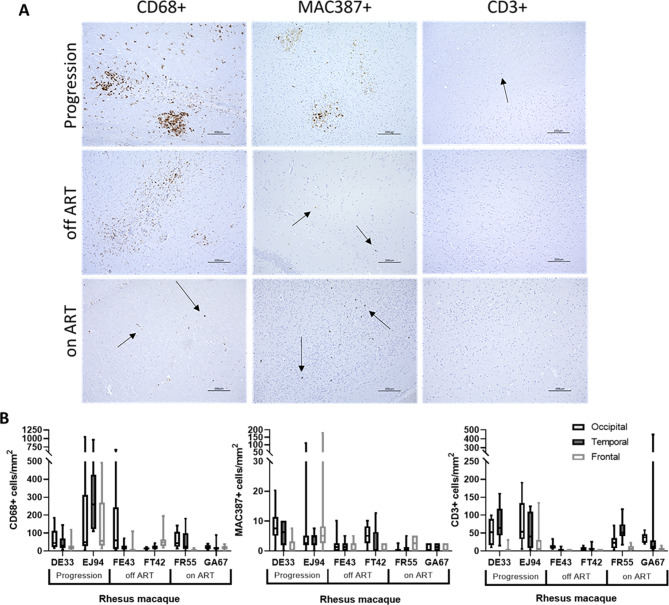
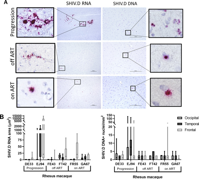
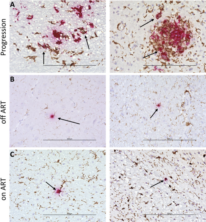
A biologically relevant non-human primate (NHP) model of HIV persistence in the central nervous system (CNS) is necessary. Most current NHP/SIV models of HIV infection fail to recapitulate viral persistence in the CNS without encephalitis or fail to employ viruses that authentically represent the ongoing HIV-1 pandemic. Here, we demonstrate viral replication in the brain and neuropathogenesis after combination antiretroviral therapy (ART) in rhesus macaques (RMs) using novel macrophage-tropic transmitted/founder (TF) simian-human immunodeficiency virus SHIV.D.191,859 (SHIV.D). Quantitative immunohistochemistry (IHC) and DNA/RNAscope in situ hybridization (ISH) were performed on three brain regions from six SHIV.D-infected RMs; two necropsied while viremic, two during analytical treatment interruptions, and two on suppressive ART. We demonstrated myeloid-mediated neuroinflammation, viral replication, and proviral DNA in the brain in all animals. These results demonstrate that TF SHIV.D models native HIV-1 CNS replication, pathogenesis, and persistence on ART in rhesus macaques.

Keywords: HIV; NeuroHIV; Non-human primates; Persistence; SHIV.


Conflict of interest statement

Dr. Burdo is a member of the Scientific Advisory Board and holds equity in Excision BioTherapeutics (unrelated to this study). All other authors declare no conflicts of interest.

Figures

Fig. 1
Fig. 1
Quantitative immunohistochemistry of immune cell markers in TF SHIV.D RM brain tissue. Immunohistochemistry (IHC) staining of CD68+, MAC387+, and CD3 + cells in brain tissue of progression, off-ART, and ART suppressed RMs (A). Representative images from occipital, temporal, and frontal lobes at 10X magnification are shown. Scale bar = 200 μm. Target cells were quantified using Keyence BZ-X700 Microscope and accompanying Batch Analysis Software to determine the average number of IHC stained cells from 10 nonoverlapping 20X images per brain section of each RM (B). 20X frame area = 393,880 μm². Box and whisker plots are presented as mean values of counts ± quartiles, with whiskers representing the range
Fig. 2
Fig. 2
Quantitative viral RNA and DNA in situ hybridization in TF SHIV.D RM brain tissue. RNAscope (left) and DNAscope (right) in situ hybridization (ISH) of replicating viral RNA and proviral DNA in brain tissue of progression, off-ART, and ART suppressed RM (A). Representative images from occipital, temporal, and frontal lobes are shown at 10X (RNA) and 20X (DNA) magnification. Inserts = 40X magnification. Scale bar = 200 μm. The area of SHIV.D RNA positive signal (µm2) or the number of nuclei containing proviral DNA/mm2 were quantified using Keyence BZ-X700 Microscope and accompanying Batch Analysis Software to determine the average area of positive signal (RNA) or the mean number of SHIV.D DNA-containing nuclei (DNA) from 10 nonoverlapping 20X images per brain section of each RM (B). 20X frame area = 393,880 μm². Box and whisker plots are presented as mean values of counts ± quartiles, with whiskers representing the range
Fig. 3
Fig. 3
TF SHIV.D replicates and persists in myeloid cells. Dual RNAscope ISH (red) and IBA1 IHC (brown) reveal RNA/IBA1 + microglia/macrophage co-localization (arrows) in viremic RM EJ94 (A) and at low levels during ATI in off ART RM FE43 and FT42 (B) and ART suppression in on-ART RM GA67 and FR55 (C). Representative images are 40X magnification. Scale bar = 200 μm

References

    1. Castro-Gonzalez S, Colomer-Lluch M, Serra-Moreno R. Barriers for HIV cure: the Latent Reservoir. AIDS Res Hum Retroviruses. 2018;34(9):739–59. doi: 10.1089/aid.2018.0118. - DOI - PMC - PubMed
    1. Koenig S, Gendelman HE, Orenstein JM, Canto MCD, Pezeshkpour GH, Yungbluth M, Janotta F, Aksamit A, Martin MA, Fauci AS. Detection of AIDS Virus in Macrophages in Brain tissue from AIDS patients with Encephalopathy. Science; 1986. - PubMed
    1. Coleman CM, Wu L. HIV interactions with monocytes and dendritic cells: viral latency and reservoirs. Retrovirology. 2009;6(1):51. doi: 10.1186/1742-4690-6-51. - DOI - PMC - PubMed
    1. Kumar A, Abbas W, Herbein G. HIV-1 latency in Monocytes/Macrophages. Viruses. 2014;6(4):1837–60. doi: 10.3390/v6041837. - DOI - PMC - PubMed
    1. Moretti S, Virtuoso S, Sernicola L, Farcomeni S, Maggiorella MT, Borsetti A. Advances in SIV/SHIV Non-Human Primate Models of NeuroAIDS. Pathogens 2021, 10(8). - PMC - PubMed

Publication types