

Applications of Laser Speckle Contrast Imaging Technology in Dermatology
- PMID: 37564105
- PMCID: PMC10410171
- DOI: 10.1016/j.xjidi.2023.100187
Applications of Laser Speckle Contrast Imaging Technology in Dermatology
Abstract
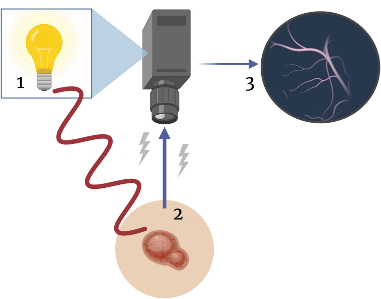
Laser speckle contrast imaging or laser speckle imaging (LSI) is a noninvasive imaging technology that can detect areas of dynamic perfusion or vascular flow. Thus, LSI has shown increasing diagnostic utility in various pathologies and has been employed for intraoperative, postoperative, and long-term monitoring in many medical specialties. Recently, LSI has gained traction in clinical dermatology because it can be effective in the assessment of pathologies that are associated with increased perfusion and hypervascularity compared with that of normal tissue. To date, LSI has been found to be highly accurate in monitoring skin graft reperfusion, determining the severity of burns, evaluating neurosurgical revascularization, assessing persistent perfusion in capillary malformations after laser therapy, and differentiating malignant and benign skin lesions. LSI affords the advantage of noninvasively assessing lesions before more invasive methods of diagnosis, such as tissue biopsy, while remaining inexpensive and exhibiting no adverse events to date. However, potential obstacles to its clinical use include tissue movement artifact, primarily qualitative data, and unclear impact on clinical practice given the lack of superiority data compared with the current standard-of-care diagnostic methods. In this review, we discuss the clinical applications of LSI in dermatology for use in the diagnosis and monitoring of vascular, neoplastic, and inflammatory skin conditions.
© 2023 The Authors.
Figures
Similar articles
-
Correcting for motion artifact in handheld laser speckle images.J Biomed Opt. 2018 Mar;23(3):1-7. doi: 10.1117/1.JBO.23.3.036006. J Biomed Opt. 2018. PMID: 29546735 Free PMC article.
-
Quantitative long-term measurements of burns in a rat model using Spatial Frequency Domain Imaging (SFDI) and Laser Speckle Imaging (LSI).Lasers Surg Med. 2017 Mar;49(3):293-304. doi: 10.1002/lsm.22647. Epub 2017 Feb 21. Lasers Surg Med. 2017. PMID: 28220508 Free PMC article.
-
Assessing multimodal optical imaging of perfusion in burn wounds.Burns. 2022 Jun;48(4):799-807. doi: 10.1016/j.burns.2021.08.026. Epub 2021 Sep 4. Burns. 2022. PMID: 34696954
-
Advances in laser speckle imaging: From qualitative to quantitative hemodynamic assessment.J Biophotonics. 2024 Jan;17(1):e202300126. doi: 10.1002/jbio.202300126. Epub 2023 Oct 3. J Biophotonics. 2024. PMID: 37545037 Review.
-
Clinical applications of laser speckle contrast imaging: a review.J Biomed Opt. 2019 Aug;24(8):1-11. doi: 10.1117/1.JBO.24.8.080901. J Biomed Opt. 2019. PMID: 31385481 Free PMC article. Review.
Cited by
-
Exploring the efficacy of laser speckle contrast imaging in the stratified diagnosis of rosacea: a quantitative analysis of facial blood flow dynamics across varied regions.Front Immunol. 2024 Aug 23;15:1419005. doi: 10.3389/fimmu.2024.1419005. eCollection 2024. Front Immunol. 2024. PMID: 39247187 Free PMC article.
-
Exploring the Role of Perfusion in Skin Graft Viability on the Scalp and Lower Limb: An Analysis of Graft Bed, Margin, and Donor Skin Using Laser Speckle.J Clin Med. 2024 Dec 16;13(24):7671. doi: 10.3390/jcm13247671. J Clin Med. 2024. PMID: 39768595 Free PMC article.
-
Noninvasive monitoring of vascular alterations in mice with acute lower limb ischemia using multimodal photoacoustic imaging.Bioeng Transl Med. 2025 Feb 17;10(4):e70005. doi: 10.1002/btm2.70005. eCollection 2025 Jul. Bioeng Transl Med. 2025. PMID: 40708973 Free PMC article.
-
Potential Utility of Laser Speckle Contrast Imaging to Detect Early Microcirculatory Changes Associated With Erythema Nodosum in Crohn's Disease: A Case Report.Inflamm Bowel Dis. 2025 May 12;31(5):1471-1472. doi: 10.1093/ibd/izae284. Inflamm Bowel Dis. 2025. PMID: 39607856
-
Precision diagnosis of burn injuries using imaging and predictive modeling for clinical applications.Sci Rep. 2025 Mar 4;15(1):7604. doi: 10.1038/s41598-025-92096-4. Sci Rep. 2025. PMID: 40038450 Free PMC article.
References
-
- Agnew K.L., Gilchrest B.A., Bunker C.B. Health Press Limited; Abingdon, United Kingdom: 2005. Fast facts. Skin Cancer.
-
- Aminfar A., Davoodzadeh N., Aguilar G., Princevac M. Application of optical flow algorithms to laser speckle imaging. Microvasc Res. 2019;122:52–59. - PubMed
-
- Bamps D., Macours L., Buntinx L., de Hoon J. Laser speckle contrast imaging, the future DBF imaging technique for TRP target engagement biomarker assays. Microvasc Res. 2020;129 - PubMed
Publication types
LinkOut - more resources
Full Text Sources
