Endoscopic features of low-grade dysplastic Barrett's
- PMID: 37564334
- PMCID: PMC10411114
- DOI: 10.1055/a-2102-7726
Endoscopic features of low-grade dysplastic Barrett's
Abstract
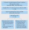
Background and study aims Barrett's esophagus (BE) with low-grade dysplasia (LGD) is considered usually endoscopically invisible and the endoscopic features are not well described. This study aimed to: 1) evaluate the frequency of visible BE-LGD; 2) compare rates of BE-LGD detection in the community versus a Barrett's referral unit (BRU); and 3) evaluate the endoscopic features of BE-LGD. Patients and methods This was a retrospective analysis of a prospectively observed cohort of 497 patients referred to a BRU with dysplastic BE between 2008 and 2022. BE-LGD was defined as confirmation of LGD by expert gastrointestinal pathologist(s). Endoscopy reports, images and histology reports were reviewed to evaluate the frequency of endoscopically identifiable BE-LGD and their endoscopic features. Results A total of 135 patients (27.2%) had confirmed BE-LGD, of whom 15 (11.1%) had visible LGD identified in the community. After BRU assessment, visible LGD was detected in 68 patients (50.4%). Three phenotypes were observed: (A) Non-visible LGD; (B) Elevated (Paris 0-IIa) lesions; and (C) Flat (Paris 0-IIb) lesions with abnormal mucosal and/or vascular patterns with clear demarcation from regular flat BE. The majority (64.7%) of visible LGD was flat lesions with abnormal mucosal and vascular patterns. Endoscopic detection of BE-LGD increased over time (38.7% (2009-2012) vs. 54.3% (2018-2022)). Conclusions In this cohort, 50.4% of true BE-LGD was endoscopically visible, with increased recognition endoscopically over time and a higher rate of visible LGD detected at a BRU when compared with the community. BRU assessment of BE-LGD remains crucial; however, improving endoscopy surveillance quality in the community is equally important.
Keywords: Barrett's and adenocarcinoma; Diagnosis and imaging (inc chromoendoscopy, NBI, iSCAN, FICE, CLE); Endoscopic resection (ESD, EMRc, ...); Endoscopy Upper GI Tract; Image and data processing, documentatiton; Quality and logistical aspects.
The Author(s). This is an open access article published by Thieme under the terms of the Creative Commons Attribution-NonDerivative-NonCommercial-License, permitting copying and reproduction so long as the original work is given appropriate credit. Contents may not be used for commercial purposes, or adapted, remixed, transformed or built upon. (https://creativecommons.org/licenses/by-nc-nd/4.0/).
Conflict of interest statement
Conflict of Interest The authors declare that they have no conflict of interest.
Figures


Comment in
-
Low-grade dysplasia on Barrett's esophagus: visible or not ?Endosc Int Open. 2023 Sep 1;11(9):E816-E817. doi: 10.1055/a-2145-5564. eCollection 2023 Sep. Endosc Int Open. 2023. PMID: 37664789 Free PMC article. No abstract available.
References
-
- Solanky D, Krishnamoorthi R, Crews N et al. Barrett oesophagus length, nodularity, and low-grade dysplasia are predictive of progression to oesophageal adenocarcinoma. J Clin Gastroenterol. 2019;53:361–365. - PubMed
-
- Hameeteman W, Tytgat GN, Houthoff HJ et al. Barrett's oesophagus: development of dysplasia and adenocarcinoma. Gastroenterology. 1989;96:1 249–1256. - PubMed
-
- Wani S, Muthusamy VR, Shaheen NJ et al. Development of quality indicators for endoscopic eradication therapies in Barrett's oesophagus: the TREAT-BE (Treatment with Resection and Endoscopic Ablation Techniques for Barrett's oesophagus) Consortium. Gastrointest Endosc. 2017;86:1–17000. - PubMed
-
- Duits LC, Phoa KN, Curvers WL et al. Barrett's oesophagus patients with low-grade dysplasia can be accurately risk-stratified after histological review by an expert pathology panel. Gut. 2015;64:700–706. - PubMed
LinkOut - more resources
Full Text Sources
Miscellaneous

