. 2022 Apr 22:3:e6.
doi: 10.1017/qrd.2022.1. eCollection 2022.

Pulmonary surfactant and COVID-19: A new synthesis

Affiliations

Pulmonary surfactant and COVID-19: A new synthesis

Barry Ninham et al. QRB Discov. .

Abstract

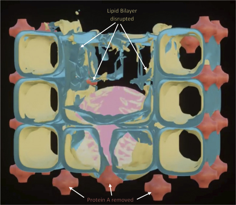
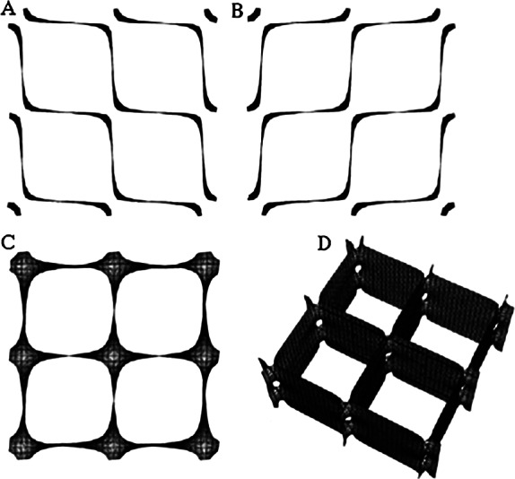
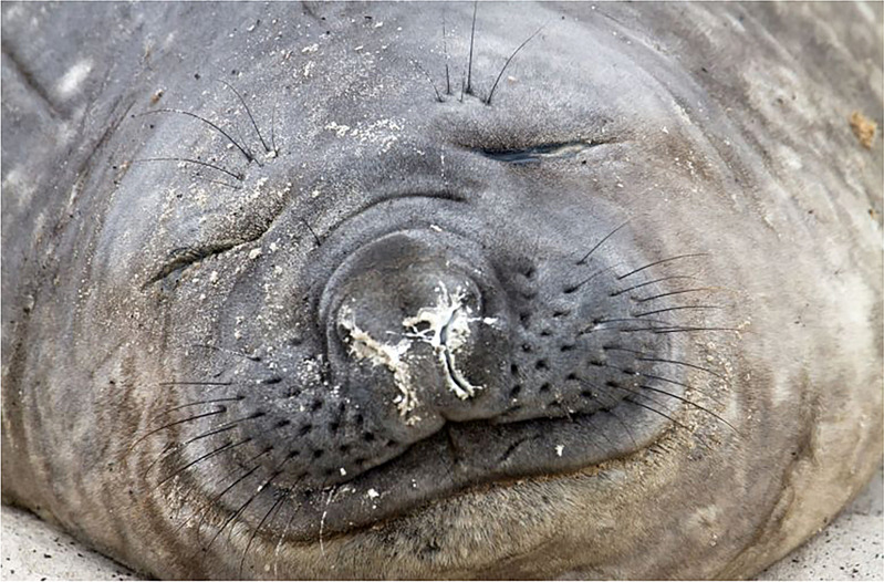
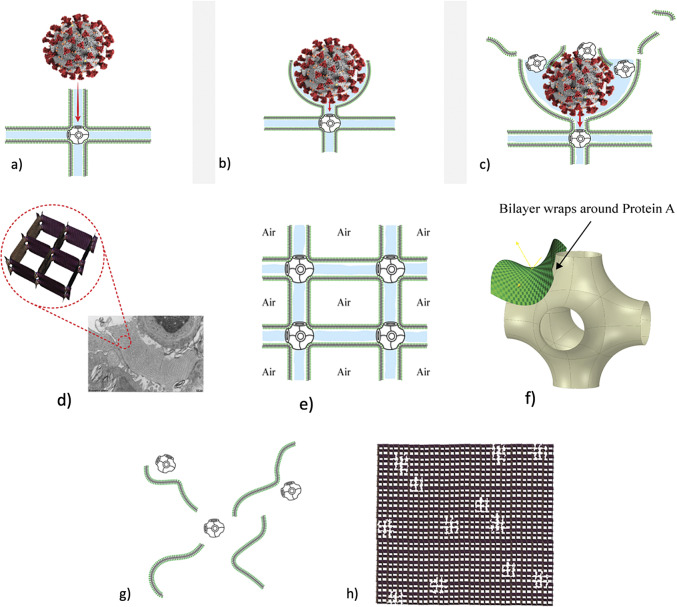
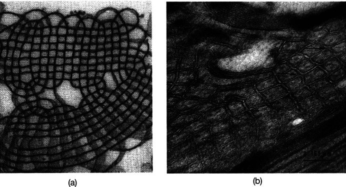
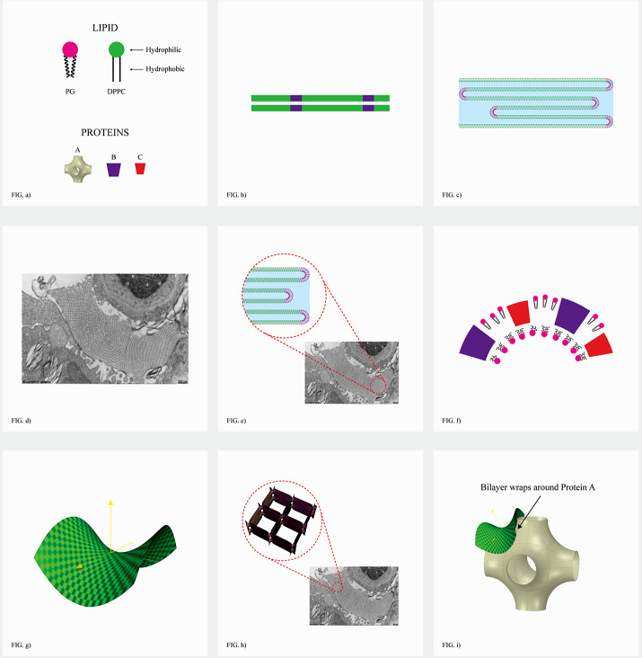
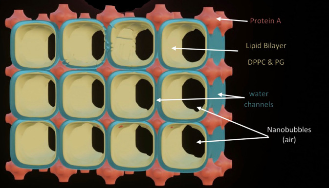
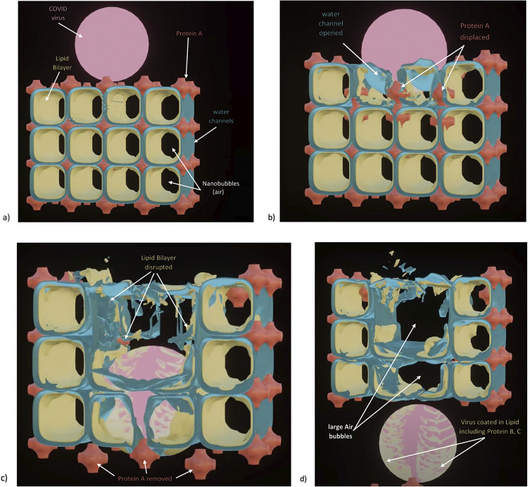
Chapter 1: COVID-19 pathogenesis poses paradoxes difficult to explain with traditional physiology. For instance, since type II pneumocytes are considered the primary cellular target of SARS-CoV-2; as these produce pulmonary surfactant (PS), the possibility that insufficient PS plays a role in COVID-19 pathogenesis has been raised. However, the opposite of predicted high alveolar surface tension is found in many early COVID-19 patients: paradoxically normal lung volumes and high compliance occur, with profound hypoxemia. That 'COVID anomaly' was quickly rationalised by invoking traditional vascular mechanisms-mainly because of surprisingly preserved alveolar surface in early hypoxemic cases. However, that quick rejection of alveolar damage only occurred because the actual mechanism of gas exchange has long been presumed to be non-problematic, due to diffusion through the alveolar surface. On the contrary, we provide physical chemical evidence that gas exchange occurs by an process of expansion and contraction of the three-dimensional structures of PS and its associated proteins. This view explains anomalous observations from the level of cryo-TEM to whole individuals. It encompasses results from premature infants to the deepest diving seals. Once understood, the COVID anomaly dissolves and is straightforwardly explained as covert viral damage to the 3D structure of PS, with direct treatment implications. As a natural experiment, the SARS-CoV-2 virus itself has helped us to simplify and clarify not only the nature of dyspnea and its relationship to pulmonary compliance, but also the fine detail of the PS including such features as water channels which had heretofore been entirely unexpected.

Chapter 2: For a long time, physical, colloid and surface chemistry have not intersected with physiology and cell biology as much as we might have hoped. The reasons are starting to become clear. The discipline of physical chemistry suffered from serious unrecognised omissions that rendered it ineffective. These foundational defects included omission of specific ion molecular forces and hydration effects. The discipline lacked a predictive theory of self-assembly of lipids and proteins. Worse, theory omitted any role for dissolved gases, O2, N2, CO2, and their existence as stable nanobubbles above physiological salt concentration. Recent developments have gone some way to explaining the foam-like lung surfactant structures and function. It delivers O2/N2 as nanobubbles, and efflux of CO2, and H2O nanobubbles at the alveolar surface. Knowledge of pulmonary surfactant structure allows an explanation of the mechanism of corona virus entry, and differences in infectivity of different variants. CO2 nanobubbles, resulting from metabolism passing through the molecular frit provided by the glycocalyx of venous tissue, forms the previously unexplained foam which is the endothelial surface layer. CO2 nanobubbles turn out to be lethal to viruses, providing a plausible explanation for the origin of 'Long COVID'. Circulating nanobubbles, stable above physiological 0.17 M salt drive various enzyme-like activities and chemical reactions. Awareness of the microstructure of Pulmonary Surfactant and that nanobubbles of (O2/N2) and CO2 are integral to respiratory and circulatory physiology provides new insights to the COVID-19 and other pathogen activity.

Keywords: COVID; Gas Exchange; Lung Surfactant; Pulmonary Surfactant; nano-bubbles; physical chemistry.


Figures

None
Graphical abstract
Fig. 1.
Fig. 1.
Two-dimensional (2D) cryo-TEM models of actual pulmonary surfactant ex vivo and calculated 3D structure on full molecular ‘inspiration’. (ac) are sections of pulmonary surfactant (PS) ex vivo revealed by cryo-TEM studies of freshly opened alveolar surfaces of rabbit lungs, by transfer of the surface layer to the grid and immediately freezing the structure without ice crystal formation. The imaged pattern is seen in two adjacent cross-sections (a and b), and then these cross-sections overlap (c). Seeing this cubic lattice-like appearance came as a welcome surprise to Larsson et al. (1999), as it had precisely the dimensions of the lattice-like structure often seen in traditional EM known as ‘tubular myelin’ (TM). The finding that the cubic dimensions (and interbilayer distance) are approximately 40 nm in the PS found by cryo-TEM and what had been known as TM strongly suggested that they were one in the same structure, albeit slightly different microanatomic positions. This observation implied that TM is not merely a highly-ordered storage depot for PS lipids, but the O2-containing contracted state of PS at the alveolar surface, and perhaps subjacent to it (although the possibility exists that traditional EM processing methods artefactually ‘knock’ the PS at the surface to a deeper position below the alveolar surface than it normally occupies, as conceded by Jastrow himself, personal communication with BR, 10/12/2021). Image (d) is the structure calculated using Mathematica software program and adopting the nodal surface approximation, showing the postulated non-Euclidean 3D structure of pulmonary surfactant on full inspiration known as ‘crossed layers of parallels’ or ‘CLP’ (Reproduced from Andersson et al., ; Larsson and Larsson, 2014), obtaining permission of Springer Copyright 1999 and 2002. The corners of the cubes are occupied by surfactant protein A (SP-A), which holds the corners together in Lego-like fashion. The expanded and contracted states of PS are explained in more detail in Box 2, and in the second chapter.
Fig. 2.
Fig. 2.
Copious production of pulmonary surfactant in Elephant Seals. This is a photograph of an elephant seal sleeping on the beach. Note that the whitish material collecting around the external nares is not mucus, but pulmonary surfactant. Weddell seals are also known to cough or sneeze up excess pulmonary surfactant when they surface. This fits with our hypothesis that deep-diving seals switch to a completely hydrostatic system for delivering oxygen to tissues, where O2 is stored in the surfactant as nanobubbles during the dive and diffuses to all tissues at depth (as documented in 1959 in swine experiments where erythrocytes were depleted, and oxygen delivered under hyperbaric conditions). It has been documented in vitro that type II pneumocytes of seals can produce surfactant at high pressures of deep dives, and likely produce it abundantly.
Fig. 3.
Fig. 3.
How SARS-CoV-2 disrupts the 3D structure of pulmonary surfactant by breaking open its corners. (a) Analysis of the crisscross pattern often seen in electron micrographs of the alveolar surface reveals a 3D structure which is pulmonary surfactant (PS), shown in its full open configuration on inspiration. (b) A top-down view of the 3D structure of PS shows within its corners the hexagonal surfactant protein A (SP-A) shown in white against blue water channels, which are formed inside the polar head groups of the lipid bilayer of DPCC which comprises the main phospholipid (90%) forming the walls of the 3D structure of PS. (c) A more detailed analysis of an individual corner of PS reveals an enlarged SP-A with a single green covering of phosphatidyl glycerol (PG) which comprises 10% of the phospholipid of PS, the hydrophilicity of which allows it to have hydration compatibility with the hydrophilic surface of SP-A. (d–f) SARS-CoV-2 is shown entering a water channel of PS and moving towards a SP-A-containing corner, which it ultimately disrupts, freeing SP-A into circulation (g), and destroying the overall 3D structure of PS (h).
Fig. 4.
Fig. 4.
Schematic of bubble–bubble interaction experiment with addition of salts and sugars (see Craig et al., a, b; Nylander et al., ; Kékicheff and Ninham, ; Henry and Craig, ; Craig and Henry, 2010).
Fig. 5.
Fig. 5.
Cartoon of the problem of restriction enzymes reproduced (Kim et al., 2001).
Fig. 6.
Fig. 6.
Efficiency of a standard restriction enzyme in cutting DNA as a function of salt and salt type.
Fig. 7.
Fig. 7.
Asymmetry of the curved bilayer of a vesicle.
Fig. 8.
Fig. 8.
With multilayered liposomes, the interior collapses to a bicontinuous phase of cubic symmetry with separated channels. Mitochondria are typical examples of these ubiquitous cell organelles.
Fig. 9.
Fig. 9.
Image of cubic phase of phospholipids courtesy of Stephen Hyde.
Fig. 10.
Fig. 10.
Schematic of the 2 different lung surfactant phases (expiration, inhalation) from Perez-Gil (2008).
Fig. 11.
Fig. 11.
TEM of lung surfactant surrounding capillary. Note cubes containing O2/N2 (cf. Fig. 12). The sides of the cubes are in fact two facing bilayers separated by channels of water (Courtesy of Holger Jastrow, http://www.drjastrow.de/Demo/Lung.jpg; tubular myelin (rat)).
Fig. 12.
Fig. 12.
(a) TEM of foetal rat lung (Young et al., ; Sanderson and Vatter, 1997) and (b) TEM of rat lung (Sanderson and Vatter, ; Andersson et al.,1999) showing tubular myelin in cubic phase of lung surfactant consistent with Fig. 10 (lower) and Fig. 11 (left side) also found in Andersson et al. (1999).
Fig. 13.
Fig. 13.
The nuts and bolts of lung surfactant. SP-D is small, but self assembles into complex roles (see section ‘Lung surfactant D and other pathologies’). (a) Nuts and bolts: Membrane forming lipids (double chained) (90%) DPPC [di palmitoyl (C16) 2 diphosphotidyl choline] and 10% PG [phosphatidyl glycerol], plus cholesterol (not depicted)-membrane stiffener, and four lung surfactant proteins, SP-A large and SP-D small are very hydrophilic. SP Proteins B and C are very hydrophobic. SP-D self assembles into complex structures and has a complex role described in section ‘Lung surfactant D and other pathologies’. (b) Hydrophobic association of lipids produces membranes coloured green (DPPC) and red (PG). lLipids separate into rigid planar regions (DPPC) that repel viruses and bacteria, and softer PG regions. Such a phase separations occur in any mixture and are crucial to formation of nanostructures required. (c) Expanded lung surfactant is a single extended folded bilayer (giant vesicle) after expulsion of CO2/water nanobubbles (and virus). (Tubular myelin) Aqueous thicknesses are about 50 nm (thicknesses of usual liposomes of membrane lipids is only 3 nm). (d) Actual lung surfactant showing coexistence of both expanded (Fig. 11 RHS) and (Fig. 11 LHS) (tetragonal cubic) phase nano-compartments of O2/N2. (e) Expanded lung surfactant after delivery of gas nanobubbles. (f) The curved bilayers at the corners of the cubic gas containers. (f,g) are formed of PG (hydrophilic head group) and the hydrophobic SP-B and C that allow necessary (green) saddle-shaped joining region of (i) made by the pseudo ‘lego’ SP-A. (h) Image shows how the bilayers of the expanded lung surfactant phase folds down to form connected compartments of a phase of cubic symmetry containing O2/N2 nanobubbles. (i) Schematic of the topology of the large hydrophilic SP-A, showing six arms to join corners of stacks of cubes containing gas.
Fig. 14.
Fig. 14.
The double bilayer water-sandwich model.
Fig. 15.
Fig. 15.
(a)–(d) The virus opening up of the water channels. The virus is the size of 2–3 compartments ~1100 nm. This releases SP-A.
Fig. 16.
Fig. 16.
Torn sweater is analogous to the damaged PS structure preventing single bilayer to structure phase change.

Similar articles

Cited by

References

Chapter 1

    1. Abbasi S, Bhutani VK and Gerdes JS (1993) Long-term pulmonary consequences of respiratory distress syndrome in preterm infants treated with exogenous surfactant. The Journal of Pediatrics 122, 446–452. 10.1016/s0022-3476(05)83439-0. - DOI - PubMed
    1. Acharya D, Liu G and Gack MU (2020) Dysregulation of type I interferon responses in COVID-19. Nature Reviews Immunology 20, 397–398. 10.1038/s41577-020-0346-x. - DOI - PMC - PubMed
    1. Ackermann M, Verleden SE, Kuehnel M, Haverich A, Welte T, Laenger F, Vanstapel A, Werlein C, Stark H, Tzankov A, Li WW, Li VW, Mentzer SJ and Jonigk D (2020) Pulmonary vascular endothelialitis, thrombosis, and angiogenesis in COVID-19. The New England Journal of Medicine 383, 120–128. 10.1056/NEJMoa2015432. - DOI - PMC - PubMed
    1. Ahmetaj-Shala B, Peacock P, Baillon L, Swann OC, Gashaw H, Barclay WS and Mitchell JA (2021) Resistance of endothelial cells to SARS-CoV-2 infection in vitro. bioRxiv. 10.1101/2020.11.08.372581. - DOI
    1. Alexander J and Milner AD (1995) Lung volume and pulmonary blood flow measurements following exogenous surfactant. European Journal of Pediatrics 154, 392–397. 10.1007/BF02072113. - DOI - PubMed

Chapter 2

The Background
    1. Lo Nostro P and Ninham BW (2020) After DLVO: Hans Lyklema and the keepers of the faith. Advances in Colloid and Interface Science 276, 102082. - PubMed
    1. Ninham BW (2006) The present state of molecular forces. Progress in Colloid and Polymer Science 133, 65–73. 10.1007/2882_051. - DOI
    1. Ninham BW (2017) The biological/physical sciences divide and the age of unreason. Substantia 1(1), 7–24. 10.13128/Substantia-6. - DOI
    1. Ninham BW (2019) B. V. Derjaguin and J. Theo. G. Overbeek. Their times, and ours. Substantia 3(2), 65–72. 10.13128/Substantia-637. - DOI
    1. Ninham BW and Lo Nostro P (2010) Molecular Forces and Self-Assembly: In Colloid, Nano Sciences and Biology. Cambridge: Cambridge University Press.
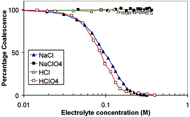
Bubble–bubble fusion in electrolytes
    1. Craig V and Henry C (2010) Inhibition of bubble coalescence by salts and sugars. In XXV International Mineral Processing Congress: IMPC 2010 Proceedings, Brisbane, Australia, ed. Conference Program Committee, CSIRO Publishing pp. 1814–1826.
    1. Henry CL and Craig VSG (2009) Inhibition of bubble coalescence by osmolytes: Sucrose, other sugars, and urea. Langmuir 25, 11406–11412. - PubMed
    1. Kékicheff P and Ninham BW (1990) The double-layer interaction of asymmetric electrolytes. Europhysics Letters 12(5), 471–477. 10.1209/0295-5075/12/5/016. - DOI
    1. Nylander T, Kékicheff P and Ninham BW (1994) The effect of solution behavior of insulin on interactions between adsorbed layers of insulin. Journal of Colloid and Interface Science 164(1), 136–150. 10.1006/jcis.1994.1152. - DOI
Classical electrolyte theory and colloid science
    1. Boström M, Craig VJS, Albion R, Williams DRM and Ninham BW (2003) Hofmeister effects in pH measurements: Role of added salt and co-ions. The Journal of Physical Chemistry. B 107(13), 2875–2878. 10.1021/jp026804d. - DOI
    1. Ninham BW, Kurihara K and Vinogradova OI (1997) Hydrophobicity, specific ion adsorption and reactivity. Colloids Surfaces A: Physicochemical and Engineering Aspects 123–124, 7–12. 10.1016/S0927-7757(96)03794-6. - DOI
    1. Salis A, Cappai L, Carucci C, Parsons DF and Monduzzi M (2020) Specific buffer effects on the intermolecular interactions among protein molecules at physiological pH. Journal of Physical Chemistry Letters 11, 6805–6811. 10.1021/acs.jpclett.0c01900. - DOI - PubMed
    1. Salis A, Pinna MC, Bilaniĉova D, Monduzzi M, Lo Nostro P and Ninham BW (2006) Specific anion effects on glass electrode pH measurements of buffer solutions: Bulk and surface phenomena. The Journal of Physical Chemistry. B 110(6), 2949–2956. 10.1021/jp0546296. - DOI - PubMed
Dissolved atmospheric gas
    1. Ninham BW and Pashley RM (2020) ‘Outline’, ‘Postscript’, & ‘cavitation’, in ‘about water: Novel technologies for the new millennium’. special issue of Substantia 4(2 Suppl.), 5–8. 10.36253/Substantia-1155. - DOI
    1. Taseidifar M, Antony J and Pashley RM (2020) Prevention of cavitation in propellers. Substantia 4(2 Suppl.), 109–117. 10.36253/Substantia-821. - DOI
Nanobubbles
    1. Alheshibri M and Craig VSJ (2018) Differentiating between nanoparticles and nanobubbles by evaluation of the compressibility and density of nanoparticles. The Journal of Physical Chemistry C 122, 21998–22007. 10.1021/acs.jpcc.8b07174. - DOI
    1. Alheshibri M and Craig VSJ (2019a) Armoured nanobubbles; ultrasound contrast agents under pressure. Journal of Colloid and Interface Science 537, 123–131. 10.1016/j.jcis.2018.10.108. - DOI - PubMed
    1. Alheshibri M and Craig VSJ (2019b) Generation of nanoparticles upon mixing ethanol and water: Nanobubbles or not? Journal of Colloid and Interface Science 542, 136–143. 10.1016/j.jcis.2019.01.134. - DOI - PubMed
    1. Alheshibri M, Jehannin M, Coleman VA and Craig VSJ (2019) Does gas supersaturation by a chemical reaction produce bulk nanobubbles? Journal of Colloid and Interface Science 554, 388–395. 10.1016/j.jcis.2019.07.016. - DOI - PubMed
    1. Bunkin NF, Kiseleva OA, Lobeyev AV, Movchan TG, Ninham BW and Vinogradova OI (1997) Effect of salts and dissolved gas on optical cavitation near hydrophobic and hydrophilic surfaces. Langmuir 13(11), 3024–3028. 10.1021/la960265k. - DOI
Carbon dioxide bubbles destroy viruses and bacteria: CO2 a saint not sinner
    1. Adams MH (1948) Surface inactivation of bacterial viruses and of proteins. The Journal of General Physiology 31(5), 417–431. - PMC - PubMed
    1. Arkill KP, Knupp C, Michel CC, Neal CR, Qvortrup K, Rostgaard J and Squire JM (2011) Similar endothelial glycocalyx structures in microvessels from a range of mammalian tissues: Evidence for a common filtering mechanism? Biophysical Journal 101(5), 1046–1056. 10.1016/j.bpj.2011.07.036. - DOI - PMC - PubMed
    1. Bunkin NF, Shkirin AV, Ninham BW, Chirikov SN, Chaikov LL, Penkov NV, Kozlov VA and Gudkov SV (2020) Shaking-induced aggregation and flotation in immunoglobulin. Dispersions: Differences between water and water−ethanol mixtures. ACS Omega 5, 14689–14701. - PMC - PubMed
    1. El-Betany AMM, Behiry E, Gumbleton M and Harding K (2020) Humidified warmed CO2 treatment therapy strategies can save lives with mitigation and suppression of SARS-CoV-2 infection: An evidence review. Frontiers in Medicine 7, 594295. 10.3389/fmed.2020.594295. - DOI - PMC - PubMed
    1. Fang Z, Wang X, Zhou L, Zhang L and Hu J (2020) Formation and stability of bulk nanobubbles by vibration. Langmuir 36, 2264–2270. 10.1021/acs.langmuir.0c00036. - DOI - PubMed
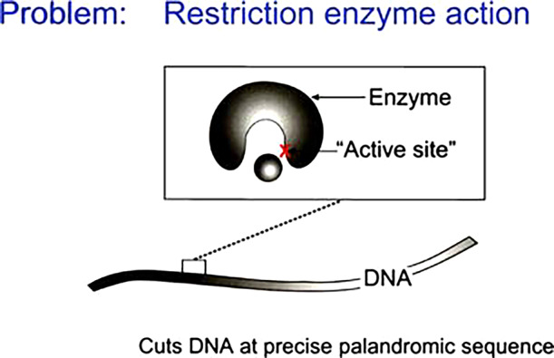
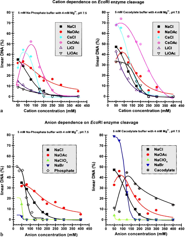
Chemical reactions, enzyme action, nanobubbles
    1. Allen M, Evans DF, Mitchell DJ and Ninham BW (1987) Interfacial tension of ionic microemulsions. Journal of Physical Chemistry 91(9), 2320–2324. 10.1021/j100293a022. - DOI
    1. Evans DF, Mitchell DJ and Ninham BW (1986) Oil, water, and surfactant: Properties and conjectured structure of simple microemulsions. The Journal of Physical Chemistry 90(13), 2817–2825. 10.1021/j100404a009. - DOI
    1. Feng B, Sosa RP, Mårtensson AKF, Jiang K, Tong A, Dorfman KD, Takahashi M, Lincoln P, Bustamante CJ, Westerlund F and Nordén B (2019) Hydrophobic catalysis and a potential biological role of DNA unstacking induced by environment effects. Proceedings of the National Academy of Sciences of the United States of America 116, 17169–17174. - PMC - PubMed
    1. Knackstedt MA and Ninham BW (1994) Model disorder media for ternary microemulsions. Physical Review E 50, 2839–2843. 10.1103/PhysRevE.50.2839. - DOI - PubMed
    1. Knackstedt MA and Ninham BW (1995) Ternary microemulsions as model disordered media. AICHE Journal 41(5), 1295–1305. 10.1002/aic.690410524. - DOI
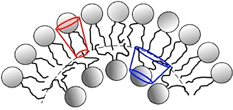
Lung surfactant structure: Self-assembly of lipids
    1. Alfredsson V, Lo Nostro P, Ninham BW and Nylander T (2021) Morphologies and structure of brain lipid membrane dispersions. Frontiers in Cell and Developmental Biology 9, 675140. 10.3389/fcell.2021.675140. - DOI - PMC - PubMed
    1. Bangham AD (1995) Surface tension in the lungs. Biophysical Journal 68, 1630–1633. https://www.ncbi.nlm.nih.gov/pmc/articles/PMC1282060/pdf/biophysj00063-0.... - PMC - PubMed
    1. Bangham D, Morley CJ and Phillips MC (1979) The physical properties of an effective lung surfactant. Biochimica et Biophysica Acta (BBA) 573(3), 552–556. 10.1016/0005-2760(79)90229-7. - DOI - PubMed
    1. Follows D, Tiberg F, Thomas RK and Larsson M (2007) Multilayers at the surface of solutions of exogenous lung surfactant: Direct observation by neutron reflection. Biochimica et Biophysica Acta 1768, 228–235. - PubMed
    1. Gershfeld NL (1989) The critical unilamellar lipid state: A perspective for membrane bilayer assembly. Biochimica et Biophysica Acta 988, 335. 10.1016/0304-4157(89)90009-9. - DOI - PubMed
The real lung surfactant – Structure and machinery
    1. Almsherqi ZA (2021) Potential role of plasmalogens in the modulation of biomembrane morphology. Frontiers in Cell and Developmental Biology 9, 673917. 10.3389/fcell.2021.673917. - DOI - PMC - PubMed
    1. Angelova A, Angelov B, Drechsler M, Bizien T, Gorshkova YE and Deng Y (2021) Plasmalogen-based liquid crystalline multiphase structures involving Docosapentaenoyl derivatives inspired by biological cubic membranes. Frontiers in Cell and Developmental Biology 9, 617984. 10.3389/fcell.2021.617984. - DOI - PMC - PubMed
    1. Ashman RB, Blanden RV, Ninham BW and Evans DF (1986) Interaction of amphiphilic aggregates with cells of the immune system. Immunology Today 7(9), 278–283. 10.1016/0167-5699(86)90010-1. - DOI - PubMed
    1. Ashman RB and Ninham BW (1985) Immunosuppressive effects of cationic vesicles. Molecular Immunology 22(5), 609–612. 10.1016/0161-5890(85)90185-3. - DOI - PubMed
    1. Ball JM, Chen S and Li W (2022) Mitochondria in cone photoreceptors act as microlenses to enhance photon delivery and confer directional sensitivity to light. Science Advances 8(9), eabn2070. 10.1126/sciadv.abn2070. - DOI - PMC - PubMed

LinkOut - more resources