. 2023 Aug 8;12(8):e1460.
doi: 10.1002/cti2.1460. eCollection 2023.

Assessment of antibodies in the upper and lower human respiratory tract at steady state and after respiratory viral infection

Affiliations

Assessment of antibodies in the upper and lower human respiratory tract at steady state and after respiratory viral infection

Marios Koutsakos et al. Clin Transl Immunology. .

Abstract

Objectives: There is an increasing appreciation for the need to study mucosal antibody responses in humans. Our aim was to determine the utility of different types of samples from the human respiratory tract, specifically nasopharyngeal (NP) swabs obtained for diagnostic purposes and bronchoalveolar lavage (BAL) obtained in outpatient and inpatient settings.

Methods: We analysed antibody levels in plasma and NP swabs from 67 individuals with acute influenza as well as plasma and BAL from individuals undergoing bronchoscopy, including five control subjects as well as seven moderately and seven severely ill subjects with a respiratory viral infection. Levels of α2-macroglobulin were determined in BAL and plasma to assess plasma exudation.

Results: IgG and IgA were readily detectable in BAL and NP swabs, albeit at different ratios, while IgM levels were low. The total amount of antibody recovered from NP swabs varied greatly between study participants. Accordingly, the levels of influenza HA-specific antibodies varied, and individuals with lower amounts of total Ig in NP swabs had undetectable levels of HA-specific Ig. Similarly, the total amount of antibody recovered from BAL varied between study participants. However, severely ill patients showed evidence of increased plasma exudation, which may confound analysis of their BAL samples for mucosal antibodies.

Conclusion: Nasopharyngeal swabs collected for diagnostic purposes may have utility in assessing antibodies from the human nasal mucosa, but variability in sampling should be accounted for. BAL samples can be utilised to study antibodies from the lower respiratory tract, but the possibility of plasma exudation should be excluded.

Keywords: BAL; antibodies; mucosal immunity; nasopharyngeal swab.


Conflict of interest statement

The Ellebedy laboratory has received funding from Moderna, Emergent BioSolutions and AbbVie, which are unrelated to the data presented in the current study. JST has received consulting fees from Curevac. AHE has received consulting and speaking fees from InBios International, Inc, Fimbrion Therapeutics, RGAX, Mubadala Investment Company, Moderna, Pfizer, GSK, Danaher, Third Rock Ventures, Goldman Sachs and Morgan Stanley and is the founder of ImmuneBio Consulting. JST and AHE are recipients of a licensing agreement with Abbvie that is unrelated to the data presented in the current study. Other authors declare no conflicts of interest.

Figures
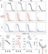
Figure 1
Figure 1
Detection of antibodies in BAL samples and nasopharyngeal swabs. (a) ELISA curves from plasma, NP swab and BAL for total Ig (heavy and light chain), IgG, IgA and IgM. Measured optical density at 490 nm and a fitted sigmoidal 4 parameter logistic curves are shown. Each curve represents a different subject. The dotted horizontal line represents the cut‐off value for endpoint titre calculations (3× background signal). (b) Interpolated concentrations of IgG, IgA and IgM for paired plasma, NP swab and BAL samples. (c) Ratios of IgG and IgA concentrations in paired samples. Samples from control subjects were used for this figure (n = 8). Statistical significance was determined by a Friedman's test with Dunn's correction for multiple comparisons.
Figure 2
Figure 2
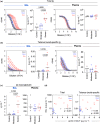
Variability in sampling of antibodies from nasopharyngeal swabs. (a) Concentrations of total Ig and IgA in plasma and NP swabs from EDFLU subjects (n = 67). Each datapoint represents a different subject and the line shows the median. (b) Concentrations of total Ig and IgA in plasma and NP swabs from EDFLU subjects grouped by sex (male n = 30, female n = 37). Median and 95% confidence intervals are shown. Statistical significance was determined by a two‐way ANOVA with Sidak's correction for multiple comparisons. (c) Correlation between concentrations of total Ig and IgA in plasma or NP swabs and age. Spearman's r coefficient and P‐value are shown for statistically significant comparisons. (d) Concentrations of total Ig and IgA in NP swabs from EDFLU subjects grouped based on disease severity (moderate n = 43, severe n = 24) and control subjects (n = 6). Statistical significance was assessed by a Kruskal–Wallis test with Dunn's correction for multiple comparisons. (e) Concentrations of HA‐specific Ig and IgA in plasma and NP swabs from EDFLU subjects (n = 67). Dotted horizontal line represents the limit of detection. Samples with undetectable levels of HA‐specific antibodies were imputed as 0.001 μg mL−1. (f) Concentrations of total Ig and IgA in plasma and NP swabs from EDFLU subjects grouped based on whether HA‐specific antibodies were detectable (n = 50 for Ig, n = 44 for IgA) or not (n = 17 for Ig, n = 23 for IgA). Statistical significance was assessed by a Mann–Whitney test.
Figure 3
Figure 3
Increased exudation and plasma leakage in BAL samples from critically ill patients. (a) ELISA curves and interpolated concentrations of total Ig for BAL and plasma according to disease severity. (b) ELISA curves and interpolated concentrations of tetanus toxoid‐specific Ig for BAL and plasma according to disease severity. (c) Concentrations of α2‐macroglobulin in BAL and plasma according to disease severity. (d) Correlation between concentrations of α2‐macroglobulin in BAL and concentrations of total and tetanus toxoid‐specific Ig in BAL. In all panels of this figure, statistical significance was assessed by a Kruskal–Wallis test with Dunn's correction for multiple comparisons (n = 5 control subjects, n = 8 samples from 7 moderately ill subjects, n = 7 severely ill subjects).

References

    1. Khoury DS, Cromer D, Reynaldi A et al. Neutralizing antibody levels are highly predictive of immune protection from symptomatic SARS‐CoV‐2 infection. Nat Med 2021; 27: 1205–1211. - PubMed
    1. Sheikh‐Mohamed S, Sanders EC, Gommerman JL, Tal MC. Guardians of the oral and nasopharyngeal galaxy: IgA and protection against SARS‐CoV‐2 infection. Immunol Rev 2022; 309: 75–85. - PMC - PubMed
    1. Gould VMW, Francis JN, Anderson KJ, Georges B, Cope AV, Tregoning JS. Nasal IgA provides protection against human influenza challenge in volunteers with low serum influenza antibody titre. Front Microbiol 2017; 8: 900. - PMC - PubMed
    1. Ravichandran S, Grubbs G, Tang J et al. Systemic and mucosal immune profiling in asymptomatic and symptomatic SARS‐CoV‐2‐infected individuals reveal unlinked immune signatures. Sci Adv 2021; 7: eabi6533. - PMC - PubMed
    1. Planas D, Bruel T, Grzelak L et al. Sensitivity of infectious SARS‐CoV‐2 B.1.1.7 and B.1.351 variants to neutralizing antibodies. Nat Med 2021; 27: 917–924. - PubMed