. 2023 Aug;95(8):e29025.
doi: 10.1002/jmv.29025.

CREB1 activation promotes human papillomavirus oncogene expression and cervical cancer cell transformation

Affiliations

CREB1 activation promotes human papillomavirus oncogene expression and cervical cancer cell transformation

Yigen Li et al. J Med Virol. 2023 Aug.

Abstract

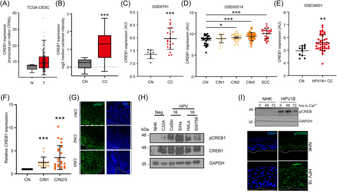
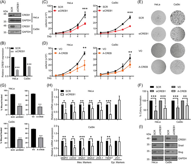
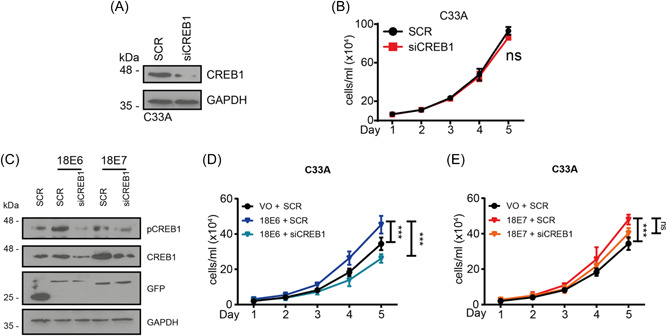
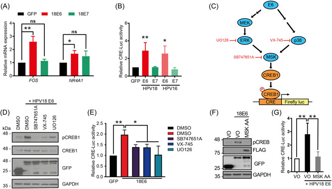
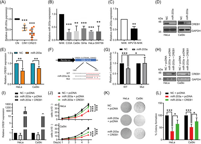
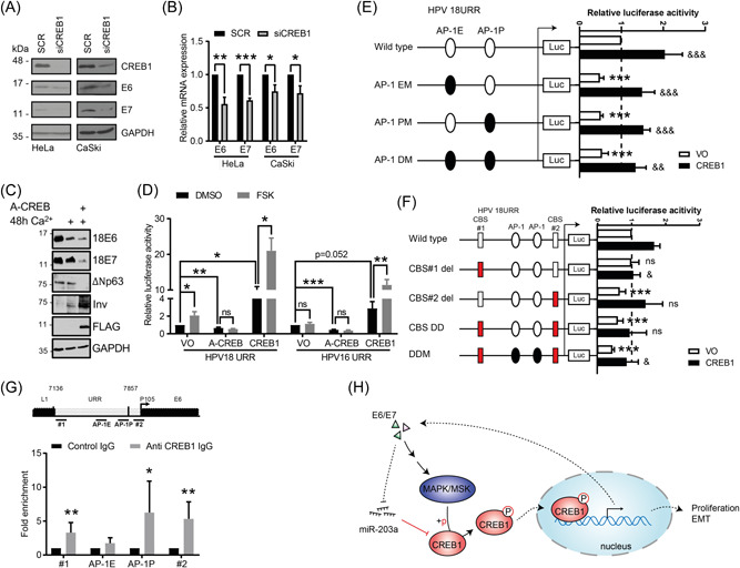
Human papillomaviruses (HPVs) infect the oral and anogenital mucosa and can cause cancer. The high-risk (HR)-HPV oncoproteins, E6 and E7, hijack cellular factors to promote cell proliferation, delay differentiation and induce genomic instability, thus predisposing infected cells to malignant transformation. cAMP response element (CRE)-binding protein 1 (CREB1) is a master transcription factor that can function as a proto-oncogene, the abnormal activity of which is associated with multiple cancers. However, little is known about the interplay between HPV and CREB1 activity in cervical cancer or the productive HPV lifecycle. We show that CREB is activated in productively infected primary keratinocytes and that CREB1 expression and phosphorylation is associated with the progression of HPV+ cervical disease. The depletion of CREB1 or inhibition of CREB1 activity results in decreased cell proliferation and reduced expression of markers of epithelial to mesenchymal transition, coupled with reduced migration in HPV+ cervical cancer cell lines. CREB1 expression is negatively regulated by the tumor suppressor microRNA, miR-203a, and CREB1 phosphorylation is controlled through the MAPK/MSK pathway. Crucially, CREB1 directly binds the viral promoter to upregulate transcription of the E6/E7 oncogenes, establishing a positive feedback loop between the HPV oncoproteins and CREB1. Our findings demonstrate the oncogenic function of CREB1 in HPV+ cervical cancer and its relationship with the HPV oncogenes.

Keywords: CREB1; E6; E7; HPV; cervical cancer; miR-203a.


Conflict of interest statement

The authors declare no conflicts of interest.

Figures

Figure 1
Figure 1
CREB1 is overexpressed in HPV containing primary keratinocytes, HPV+ cervical cancers and is associated with disease progression. (A) TCGA data analysis of CREB1 expression in cervical squamous cell carcinoma and endocervical adenocarcinoma (CESC); (B) Pyeon multicancer mRNA dataset analysis of CREB1 in cervical cancer (CC) was compared to cervical normal (CN); (C) GSE6791 dataset analysis of CREB1 expression in CC tissue was compared to CN tissue; (D) GSE63514 dataset analysis of CREB1 expression was compared among different cervical intraepithelial neoplasia (CIN) grades and cervical squamous cell carcinoma (SCC). CIN grades represent the severity of cervical disease; (E) GSE39001 dataset analysis of CREB1 expression in HPV16+ CC tissue was compared to CN tissue; (F) qPCR analysis of CREB1 in cervical cytology samples collected from healthy patients and patients with different CIN grades; (G) Representative immunostaining analysis of tissue sections from cervical lesions representing low‐grade through to high‐grade cervical disease. Sections were stained for phosphorylated CREB1 (green) and nuclei were visualized using DAPI (blue). Images were acquired using identical exposure time. (H) Western blot analysis of CREB1 in HPV+ cervical cancer cell lines compared to NHK; (I) Representative western blot analysis of normal human keratinocytes (NHK) and HPV18‐containing keratinocytes subjected to high calcium differentiation and analysed for CREB phosphorylation. GAPDH serves as loading control. Representative sections of organotypic raft cultures from NHK and HPV18‐containing keratinocytes stained with antibodies specific for phosphorylated CREB (green) and counterstained with DAPI to highlight the nuclei (blue). Images were acquired using identical exposure times. White dotted lines indicate the basal cell layer. Data shown are mean ± SD, n > 3. *p < 0.05; **p < 0.01; ***p < 0.001. CIN, cervical intraepithelial neoplasia; HPV, human papillomaviruses; mRNA, messenger ribonucleic acid.
Figure 2
Figure 2
Depletion of CREB1 inhibits the proliferation, migration and EMT of cervical cancer cells in vitro. (A) Western blot analysis of CREB1 in indicated cell lines transfected with a pool of four siRNAs targeting CREB1 (siCREB1). GAPDH loading control; (B) qPCR analysis of CREB1 in indicated cell lines transfected with siCREB1; (C, D) Cell proliferation analysed by cell growth curve in indicated cell lines transfected with siCREB1, or A‐CREB, a dominant negative inhibitor for CREB1; (E,F) Representative images for colony formation assay performed in indicated cell lines transfected with siCREB1 or A‐CREB. Quantification of (E) as shown in (F); (G) Quantification of wound closure as an evaluation for cell migration in indicated cell lines transfected with siCREB1 or A‐CREB; (H) qPCR analysis of EMT markers in indicated cell lines transfected with siCREB1; (I) Western blot analysis of EMT markers in indicated cell lines transfected with siCREB1. Data shown are mean ± SD, n ≥ 3. *p < 0.05; **p < 0.01; ***p < 0.001. EMT, epithelial to mesenchymal transition; GAPDH, glyceraldehyde 3‐phosphate dehydrogenase; siRNA, small interfering RNA.
Figure 3
Figure 3
HPV18 E6 promotes proliferation in HPV‐cervical cancer cell line C33A via CREB1. (A) Western blot analysis of CREB1 in HPV‐CC cell line, C33A, transfected with siCREB1; (B) Cell proliferation analysed by cell growth curve in HPV‐CC cell line, C33A, transfected with siCREB1; (C) Western blot analysis in C33A, co‐transfected with siCREB1 and GFP‐HPV18 E6 or E7; (D, E) Cell proliferation analysed by cell growth curve in C33A, co‐transfected with siCREB1 and HPV18 E6 or E7. Data shown are mean ± SD, n = 3. ns, not significant; ***p < 0.001. CC, cervical cancer; HPV, human papillomaviruses.
Figure 4
Figure 4
HPV E6 induces CREB1 activity via MAPK/MSK signaling. (A) qPCR analysis of CREB1‐dependent genes in C33A transfected with HPV18 E6 or E7; (B) Dual luciferase CRE‐driven luciferase reporter analysis in HEK293T transfected with HPV E6 or E7; (C) Schematic of inhibitors used for E6‐activating MAPK signaling pathways; (D, E) Western blot analysis and dual luciferase CRE‐driven luciferase reporter analysis in HEK293T transfected with HPV18 E6 and SB747651A (5 µM), VX‐745 (10 nM), or UO126 (20 µM); (F, G) Western blot analysis and dual luciferase CRE‐driven luciferase reporter analysis in C33A and HEK293T co‐transfected with HPV18 E6 and dominant negative mutant of MSK, T581A/T700A (MSK AA). Data shown are mean ± SD, n = 3. ns, no significant; *p < 0.05; **p < 0.01. CC, cervical cancer; HPV, human papillomaviruses.
Figure 5
Figure 5
miR‐203a overexpression attenuates cell proliferation by directly targeting CREB1. (A) qPCR analysis of miR‐203a in cervical cytology samples from patients with different CIN grades; (B) qPCR analysis of miR‐203a expression in CC cell lines compared to NHK; (C) qPCR analysis of miR‐203a in HPV18‐transformed NHK; (D, E) Western blot and qPCR analysis in indicated cells transfected with miR‐203a mimic; (F) Schematic of predicted miR‐203a binding sites within CREB1 3′‐UTR and point mutation introduced, fused to luciferase reporter system; (G) Dual luciferase reporter analysis of CREB1 3′‐UTR (WT and Mut) ‐controlled activity in HEK293T with overexpressing miR‐203a mimic; (H, I) Western blot and qPCR analysis of CREB1 in indicated cells co‐transfected with miR‐203a mimic and CREB1; (J–L) Cell proliferation and clonogenicity assessed by cell growth curve and colony formation assay in indicated cells co‐transfected with miR‐203a mimics and CREB1. Data shown are mean ± SD, n = 3. *p < 0.05; **p < 0.01; ***p < 0.001. CC, cervical cancer; HPV, human papillomaviruses; UTR, untranslated region.
Figure 6
Figure 6
CREB1 transcriptionally upregulates HPV oncoprotein expression by binding to the HPV URR. (A, B) Western blot and qPCR analysis of E6 and E7 expression in indicated cells transfected with siCREB1; (C) Primary HPV18 containing keratinocytes control and A‐CREB transfected and differentiated in high calcium, probed for E6 and E7 oncoproteins, p63 and involucrin as markers of proliferation/differentiation, FLAG to confirm A‐CREB expression and GAPDH as a loading control. (D) Dual luciferase HPV16/18 URR‐driven luciferase reporter analysis in HEK293T transfected with A‐CREB or CREB1, and/or stimulated with Forskolin (FSK), which promotes CREB1 activity; (E) Dual luciferase reporter analysis of HPV18 URR wild type, mutation of AP‐1 site within enhancer region (AP‐1 EM), promoter region (AP1‐PM), or both sites (AP1‐DM) activity in HEK293T with overexpressing CREB1. *, compared with wild type transfected with Vector only (VO); &, compared with the corresponding reporter construct transfected with VO. The basal transcriptional activity was set to 1; (F) Dual luciferase reporter analysis of HPV18 URR wild type, deletion of putative CREB1‐binding site 1(CBS#1 del), CBS#2 del, deletion of both sites (DD), or AP‐1 mutants with CBSs DD (DDM) activity in HEK293T with overexpressing CREB1. *, compared with wild type transfected with VO; &, compared with wild type reporter construct with overexpressing CREB1. The basal transcriptional activity was set to 1; (G) ChIP analysis of direct interaction between CREB1 with HPV18 URR. Four regions were detected: CBS#1 (#1), 7169–7313; AP‐1E, 7608–7614; AP‐1P, 7792–7798; CBS#2 (#2), 38–150; (H) Schematic of proposed model. Data shown are mean ± SD, n ≥ 3. ns, non‐significance. *p < 0.05; **p < 0.01; ***p < 0.001. HPV, human papillomaviruses; URR, upstream regulatory region.

References

    1. Schiffman MH, Bauer HM, Hoover RN, et al. Epidemiologic evidence showing that human papillomavirus infection causes most cervical intraepithelial neoplasia. J Natl Cancer Inst. 1993;85(12):958‐964. - PubMed
    1. zur Hausen H. Papillomaviruses in the causation of human cancers ‐ a brief historical account. Virology. 2009;384(2):260‐265. - PubMed
    1. de Sanjose S, Quint WG, Alemany L, et al. Human papillomavirus genotype attribution in invasive cervical cancer: a retrospective cross‐sectional worldwide study. Lancet Oncol. 2010;11(11):1048‐1056. - PubMed
    1. Ajila V, Shetty H, Babu S, Shetty V, Hegde S. Human papilloma virus associated squamous cell carcinoma of the head and neck. J Sex Transm Dis. 2015;2015:1‐5. - PMC - PubMed
    1. Watson M, Saraiya M, Wu X. Update of HPV‐associated female genital cancers in the United States, 1999‐2004. Journal of Women's Health. 2009;18(11):1731‐1738. - PubMed

Publication types

MeSH terms

Substances