Review
. 2023 Sep 15;18(18):e202300331.
doi: 10.1002/cmdc.202300331. Epub 2023 Sep 4.

Lysyl Oxidases as Targets for Cancer Therapy and Diagnostic Imaging

Affiliations
Review

Lysyl Oxidases as Targets for Cancer Therapy and Diagnostic Imaging

Reik Löser et al. ChemMedChem. .

Abstract

The understanding of the contribution of the tumour microenvironment to cancer progression and metastasis, in particular the interplay between tumour cells, fibroblasts and the extracellular matrix has grown tremendously over the last years. Lysyl oxidases are increasingly recognised as key players in this context, in addition to their function as drivers of fibrotic diseases. These insights have considerably stimulated drug discovery efforts towards lysyl oxidases as targets over the last decade. This review article summarises the biochemical and structural properties of theses enzymes. Their involvement in tumour progression and metastasis is highlighted from a biochemical point of view, taking into consideration both the extracellular and intracellular action of lysyl oxidases. More recently reported inhibitor compounds are discussed with an emphasis on their discovery, structure-activity relationships and the results of their biological characterisation. Molecular probes developed for imaging of lysyl oxidase activity are reviewed from the perspective of their detection principles, performance and biomedical applications.

Keywords: enzyme inhibitors; extracellular matrix; posttranslational modification; quinoproteins; radiotracers.


Conflict of interest statement

Conflict of Interests

The authors declare no conflict of interest.

Figures

Figure 1.
Figure 1.
Mechanism of catalysis of lysyl oxidases. Numbering of the amino acid residues is based on the human LOX isozyme. For clarity, the mononuclear copper centre is omitted in the figure.
Figure 2.
Figure 2.
Domain structure of the lysyl oxidase isozymes. PPPP = proline-rich domain. Taken with permission from Amendola et al.[30] and edited.
Figure 3.
Figure 3.
Structural model of human LOX according to Vallet et al.[24a] A) Ribbon representation, B) Surface representation with charge scale [contour colour gradients: −5.0 kT/e (red) and + 5.0 kT/e (blue)]. Created with PyMOL from data published in.[24a] The Cu2+ and Ca2+ ions are shown in ochre and grey, respectively.
Figure 4.
Figure 4.
Intra- and extracellular functions of the lysyl oxidases LOX and LOXL2 in the context of tumour progression. Further explanation is included in the text. In the case of dashed arrows, the interaction partners of the signal transduction cascades have been omitted in favour of clarity. Created with BioRender.com.
Figure 5.
Figure 5.
Functions of lysyl oxidases in the schematised process of tumour metastasis (EMT: epithelial-mesenchymal transition; MET: mesenchymal-epithelial transition). Adapted from Jones et al.[52g] with permission, Copyright © 2020, Wiley-VCH Verlag GmbH & Co. KGaA, Weinheim (Germany).
Figure 6.
Figure 6.
Newer inhibitor compounds (as of 2003) of lysyl oxidases. a All reported inhibitory activities represent IC50 values; b not reported.
Figure 7.
Figure 7.
Postulated mechanism of lysyl oxidase inhibition by hetarylamethylamine-based inhibitors.
Figure 8.
Figure 8.
Probes for fluorescence-, ELISA- and Western blot-detection of lysyl oxidases.
Figure 9.
Figure 9.
Probes for MR imaging of lysyl oxidase-generated allysine residues.
Figure 10.
Figure 10.
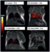
Reprinted MRT images showing the uptake of aminoxyfunctionalised Gd complex 27a and analogous unreactive methyl ether 27b as negative-control probe in the murine model of bleomycin-induced fibrosis. A) Uptake of 27a in naive mouse, showing low MR signal enhancement in healthy lungs; B) GdOA uptake in bleomycin-treated mouse, showing strong lung enhancement in 14 day bleomycin-injured mice; C) uptake of 27b in bleomycin-challenged mouse, showing low lung enhancement in 14 day bleomycin-injured mice with negative control probe and D) uptake of 27a in bleomycin-challenged mouse dosed daily for 14 days with βAPN, showing little lung enhancement, indicating an absence of allysine. Reproduced (Figure 2 in Ref. [151]) and edited (selection of subfigures, subfigure titles, compound reference numbers, caption text) from Waghorn et al.[151] with permission, Copyright © 2017, Wiley-VCH Verlag GmbH & Co. KGaA, Weinheim (Germany).
Figure 11.
Figure 11.
A) Chemical structures of probe 28a and control compound 28b for PET imaging of lysyl oxidase-generated allysine residues. B) Reprinted representative fused PET-CT (colour and gray scale, respectively) sagittal images of the head and thorax 110–120 min p.i. of (left) [68Ga]28b and (right) [68Ga]28a in sham- (SH, bottom) and bleomycin-injured (BM, top) animals; the lung is highlighted with a dashed white line. The PET signal expressed as %ID/cc, in the lung is low in both sham and BM injured mice with [68Ga]28b, but is higher in the BM injured mouse imaged with [68Ga]28a demonstrating the specificity of the latter probe for fibrogenesis. Reproduced (Figure 3 in Ref. [155]) and edited (compound reference numbers, caption text) from Wahsner et al. with permission, Copyright © 2019, American Chemical Society.
Figure 12.
Figure 12.
Substrate-based radiotracers for PET imaging of lysyl oxidases
Figure 13.
Figure 13.
Small animal PET images (maximum intensity projections) of A375 tumour-bearing NMRI (nu/nu) mice showing the distribution of [18F]29, [18F]30 and [18F]31a each after 20 min p.i. The location of the tumour tissue is indicated by an arrow. Reproduced (Figure 7 in Ref. [156]) and edited (arrangement of panels, compound reference numbers, caption text) from Kuchar et al.[156] with permission, Copyright © 2018, Kuchar et al.
Figure 14.
Figure 14.
Reprinted representative PET images after injection of [18F]31a in the absence (A) and presence (B) of β-aminopropionitrile (βAPN) in EMT-6 tumour-bearing mice at 10 min p.i. Images are shown as maximum intensity projections (MIPs) and coronal slices from the tumour region. Reproduced (Figure 10 in Ref. [116]) and edited (selection of content, caption text) from Wuest et al.[116] with permission, Copyright © 2015, Wuest et al.

Similar articles

Cited by

References

    1. Löser R, Habilitation thesis, Technische Universität Dresden (Germany), 2022. https://nbn-resolving.de/urn:nbn:de:bsz:14-qucosa2–840641.
    1. Pinnell SR, Martin GR, Proc. Natl. Acad. Sci. USA 1968, 61, 708–716. - PMC - PubMed
    1. Csiszar K, Prog. Nucleic Acid Res. Mol. Biol 2001, 70, 1–32. - PubMed
    1. Grau-Bove X, Ruiz-Trillo I, Rodriguez-Pascual F, Sci. Rep 2015, 5, 10568; - PMC - PubMed
    2. Rodriguez-Pascual F, Slatter DA, Sci. Rep 2016, 6, 37374. - PMC - PubMed
    1. Borden WT, Hoffmann R, Stuyver T, Chen B, J. Am. Chem. Soc 2017, 139, 9010–9018; - PubMed
    2. Voeikov VL, Cell. Mol. Biol. (Noisy-le-grand) 2005, 51, 663–675. - PubMed

Publication types

Substances

LinkOut - more resources