Review
. 2024 Feb;53(2):209-244.
doi: 10.1007/s00256-023-04420-6. Epub 2023 Aug 11.

SSR white paper: guidelines for utilization and performance of direct MR arthrography

Affiliations
Review

SSR white paper: guidelines for utilization and performance of direct MR arthrography

Eric Y Chang et al. Skeletal Radiol. 2024 Feb.

Erratum in

Abstract

Objective: Direct magnetic resonance arthrography (dMRA) is often considered the most accurate imaging modality for the evaluation of intra-articular structures, but utilization and performance vary widely without consensus. The purpose of this white paper is to develop consensus recommendations on behalf of the Society of Skeletal Radiology (SSR) based on published literature and expert opinion.

Materials and methods: The Standards and Guidelines Committee of the SSR identified guidelines for utilization and performance of dMRA as an important topic for study and invited all SSR members with expertise and interest to volunteer for the white paper panel. This panel was tasked with determining an outline, reviewing the relevant literature, preparing a written document summarizing the issues and controversies, and providing recommendations.

Results: Twelve SSR members with expertise in dMRA formed the ad hoc white paper authorship committee. The published literature on dMRA was reviewed and summarized, focusing on clinical indications, technical considerations, safety, imaging protocols, complications, controversies, and gaps in knowledge. Recommendations for the utilization and performance of dMRA in the shoulder, elbow, wrist, hip, knee, and ankle/foot regions were developed in group consensus.

Conclusion: Although direct MR arthrography has been previously used for a wide variety of clinical indications, the authorship panel recommends more selective application of this minimally invasive procedure. At present, direct MR arthrography remains an important procedure in the armamentarium of the musculoskeletal radiologist and is especially valuable when conventional MRI is indeterminant or results are discrepant with clinical evaluation.

Keywords: Direct MR arthrography; Labrum; Ligament; MRI; Meniscectomy; Plica; Post-operative.


Conflict of interest statement

The authors declare no competing interests.

Figures

Fig. 1
Fig. 1
Arthrographic effect from a post-traumatic hemarthrosis/effusion. Twenty-five-year-old man who fell while playing football. Axial (A), sagittal (B), and coronal (C) T2-weighted fat-suppressed conventional 3 T MR images show a tear of the inferior glenohumeral ligament complex involving the posterior band and capsule (arrows) as well as traumatic rupture of the superior cuff (arrowhead)
Fig. 2
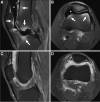
Fig. 2
“Black” contrast effect. Thirty-four-year-old man with history of meniscus surgery and persistent knee pain. Sagittal (A) and axial (B) T1-weighted fat-suppressed 3 T MR images obtained immediately after inadvertent injection of a higher concentration of gadolinium contrast shows hypointense contrast (asterisks). Notice regions of inhomogeneous fat suppression due to the strong paramagnetism of concentrated gadolinium (arrows). Sagittal (C) and axial (D) T1-weighted fat-suppressed MR images after a 3.5-h delay shows that the majority of the injectate is now hyperintense (asterisks) and there is homogeneous fat suppression
Fig. 3
Fig. 3
Intra-articular bubbles. Forty-nine-year-old woman with hip pain. Coronal (A) and sagittal (B) T1-weighted fat-suppressed 3 T MR arthrogram images show bubbles in the nondependent portion of the joint causing a dipole field pattern artifact (arrows)
Fig. 4
Fig. 4
Osteochondral lesion. Thirty-eight-year-old man with elbow pain. A Axial T2-weighted fat-suppressed conventional 1.5 T MR image shows a small osteochondral lesion at the anterior capitellum, without signs of instability (arrow). B, C Axial T1-weighted fat-suppressed 1.5 T MR arthrogram images show contrast undermining the lesion, consistent with an unstable in situ osteochondral fragment
Fig. 5
Fig. 5
Osteochondral lesion. Sixty-two-year-old woman with ankle pain. Coronal T1-weighted fat-suppressed (A), sagittal STIR (B), and sagittal T1-weighted 3 T MR arthrogram images demonstrate an osteochondral lesion at the medial aspect of the talar dome with intra-articular contrast that insinuates into the interface with the talus (arrows), consistent with instability
Fig. 6
Fig. 6
Abduction external rotation (ABER) positioning. Sixteen-year-old with history of recent shoulder dislocation. A T1-weighted fat-suppressed MR arthrogram image in the ABER position shows a tear of the anterior labrum which remains partially attached (arrow), consistent with a Perthes lesion. B On conventional axial T1-weighted fat-suppressed image, the tear is less evident (dashed arrow)
Fig. 7
Fig. 7
Anterior instability. Twenty-year-old man with recent anterior glenohumeral dislocation while playing rugby. Axial T1-weighted fat-suppressed (A) and sagittal T1-weighted (B) 1.5 T MR arthrogram images demonstrate a mixed fibrocartilaginous and osseous Bankart variant lesion with deficiency of the anterior glenoid (arrows). C Sagittal reformat image from CT scan 4 months later demonstrates the displaced osseous Bankart component (arrowhead)
Fig. 8
Fig. 8
Microinstability (type VI SLAP). Eighteen-year-old man with clinically suspected labral tear. Axial T1-weighted fat-suppressed (A) and sagittal T1-weighted (B) MR arthrogram images show a tear of the anterosuperior labrum with a fragment displaced into the anterosuperior joint recess (arrows)
Fig. 9
Fig. 9
Multidirectional instability. Eighteen-year-old man with shoulder pain. Axial T1-weighted fat-suppressed (A and B) and sagittal T2-weighted fat-suppressed (C) 1.5 T MR arthrogram images demonstrate extensive labral tearing involving both anterior and posterior aspects (arrows)
Fig. 10
Fig. 10
Chronic humeral avulsion of the glenohumeral ligament (HAGL). Twenty-seven-year-old professional pitcher with shoulder pain. A Coronal intermediate-weighted fat-suppressed conventional 1.5 T MR image shows irregularity of the inferior glenohumeral ligament (IGHL) complex (arrow) and partial-thickness tearing of the supraspinatus tendon (arrowhead). B Coronal T1-weighted fat-suppressed 1.5 T MR arthrogram image shows extra-articular contrast leakage and a thickened, retracted IGHL margin, consistent with a HAGL
Fig. 11
Fig. 11
Thrower’s shoulder. Fifteen-year-old baseball player with persistent right shoulder pain. Axial intermediate-weighted (A) and T1-weighted fat-suppressed (B) MR arthrogram images show a thickened posterior band of the inferior glenohumeral ligament at the labral insertion (arrows) with associated glenoid remodeling and retroversion. Constellation of imaging findings was consistent with the clinical diagnosis of glenohumeral internal rotation deficit (GIRD), and the patient was treated conservatively with physical therapy
Fig. 12
Fig. 12
Recurrent anterior labral tear. Twenty-six-year-old man with anterior glenohumeral dislocation 1 year following arthroscopic Bankart repair. Axial T1-weighted fat-suppressed (A) and sagittal T1-weighted (B) 1.5 T MR arthrogram images demonstrate medialized and scarred anteroinferior labroligamentous tissue (arrows), consistent with a chronic anterior labroligamentous periosteal sleeve avulsion (ALPSA). Note the anterior glenoid anchor tracks (arrowheads)
Fig. 13
Fig. 13
Thrower’s elbow. Twenty-two-year-old man with medial elbow pain and suspected ulnar collateral ligament tear. A Coronal T1-weighted fat-suppressed MR arthrogram image at the time of injury shows an ulnar collateral ligament tear at the humeral attachment (arrow) with adjacent ossicle (asterisk). B Coronal intermediate-weighted fat-suppressed conventional 3T MR image three months later shows the tear (arrow) and ossicle (asterisk) to similar advantage
Fig. 14
Fig. 14
Plica syndrome. Seventeen-year-old elite volleyball player with posterolateral elbow pain. Sagittal (A) and coronal (B) intermediate-weighted fat-suppressed conventional 1.5 T MR images demonstrate a thickened radiocapitellar plica (arrows) with adjacent bone marrow edema, consistent with the clinical diagnosis of impingement. The plica was arthroscopically resected with resolution of symptoms
Fig. 15
Fig. 15
Suspected internal derangement of the wrist. Fifty-five-year-old man with wrist pain. A Spot image of the wrist after radiocarpal joint injection shows flow of contrast into the midcarpal (small arrow) and distal radioulnar (big arrow) joints. Coronal T1-weighted 1.5 T MR arthrogram image (B) shows a tear of the lunotriquetral interosseous ligament (arrowhead) and triangular fibrocartilage (dashed arrow), which were not as clearly shown on the coronal T2-weighted fat-suppressed image (C)
Fig. 16
Fig. 16
Triangular fibrocartilage complex (TFCC) injury. A, B Coronal T1-weighted 3T MR arthrogram images show communication between the radiocarpal and distal radioulnar joints through a tear of the TFCC involving both the central disc (arrow) and ulnar attachments (arrowhead). Findings were confirmed at arthroscopy and an open foveal repair was performed
Fig. 17
Fig. 17
Postoperative wrist imaging. Thirty-eight-year-old man with wrist pain following radius fracture and surgery. A Fluoroscopic-guided direct arthrogram image shows flow of contrast from the radiocarpal joint into the distal radioulnar joint. Coronal T1-weighted 1.5 T MR arthrogram images without (B) and with (C) fat-suppression show the triangular fibrocartilage complex tear involving the central disc (arrow) and ulnar attachments (arrowhead), but the scapholunate interosseous ligament is obscured by artifact. The integrity of the scapholunate and lunotriquetral interosseous ligaments can be assumed by the lack of contrast extension into the midcarpal compartment
Fig. 18
Fig. 18
Labral tear. Forty-nine-year-old woman with hip pain. A Sagittal T2-weighted fat-suppressed conventional 3 T MR image shows a small, native joint effusion (arrow), but a labral tear is not clearly demonstrated. B Sagittal T1-weighted fat-suppressed 3 T MR arthrogram image shows contrast extending into the substance of the anterosuperior labrum (arrowhead), consistent with a tear which was subsequently confirmed at arthroscopy
Fig. 19
Fig. 19
Ligamentum teres tear. Twenty-eight-year-old ex-ballerina with chronic hip pain. Coronal T1-weighted fat-suppressed (A), coronal intermediate-weighted (B), and axial oblique T1-weighted fat-suppressed MR arthrogram images show chronic tearing and resorption of the ligamentum teres (arrowheads) as well as a torn labrum (arrows)
Fig. 20
Fig. 20
Postoperative hip imaging. Twenty-one-year-old woman with prior resections of cam and pincer lesions as well as a periacetabular osteotomy, presenting with hip pain. Axial oblique T2 fat-suppressed (A), radial T2-weighted (B), and sagittal T1-weighted fat-suppressed MR arthrogram images show a surgically confirmed recurrent tear of the labrum extending from 10 o’clock to 2 o’clock (arrowheads)
Fig. 21
Fig. 21
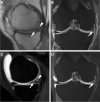
Recurrent meniscal tear. Fifty-one-year-old man with history of meniscal surgery and recurrent symptoms. Sagittal intermediate-weighted (A) and coronal intermediate-weighted fat-suppressed (B) conventional MR images show evidence of partial medial meniscectomy with linear increased signal extending to the inferior surface (arrows) and small posterior parameniscal cyst (arrowhead), consistent with a recurrent tear. Sagittal (C) and coronal (D) T1-weighted fat-suppressed MR arthrogram images demonstrate dilute gadolinium extending through the tear (arrows) and filling the parameniscal cyst (arrowhead)

Similar articles

Cited by

References

    1. Hajek PC, Baker LL, Sartoris DJ, Neumann CH, Resnick D. MR arthrography: anatomic-pathologic investigation. Radiology. 1987;163(1):141–147. doi: 10.1148/radiology.163.1.3823427. - DOI - PubMed
    1. Schweitzer ME, Brahme SK, Hodler J, Hanker GJ, Lynch TP, Flannigan BD, et al. Chronic wrist pain: spin-echo and short tau inversion recovery MR imaging and conventional and MR arthrography. Radiology. 1992;182(1):205–211. doi: 10.1148/radiology.182.1.1727283. - DOI - PubMed
    1. Palmer WE, Brown JH, Rosenthal DI. Rotator cuff: evaluation with fat-suppressed MR arthrography. Radiology. 1993;188(3):683–687. doi: 10.1148/radiology.188.3.8351333. - DOI - PubMed
    1. Hodler J, Yu JS, Goodwin D, Haghighi P, Trudell D, Resnick D. MR arthrography of the hip: improved imaging of the acetabular labrum with histologic correlation in cadavers. AJR Am J Roentgenol. 1995;165(4):887–891. doi: 10.2214/ajr.165.4.7676987. - DOI - PubMed
    1. Zanetti M, Bram J, Hodler J. Triangular fibrocartilage and intercarpal ligaments of the wrist: does MR arthrography improve standard MRI? J Magn Reson Imaging. 1997;7(3):590–594. doi: 10.1002/jmri.1880070322. - DOI - PubMed