. 2023 Aug 1;64(11):17.
doi: 10.1167/iovs.64.11.17.

PERK Inhibition Suppresses Neovascularization and Protects Neurons During Ischemia-Induced Retinopathy

Affiliations

PERK Inhibition Suppresses Neovascularization and Protects Neurons During Ischemia-Induced Retinopathy

Shuizhen Shi et al. Invest Ophthalmol Vis Sci. .

Abstract

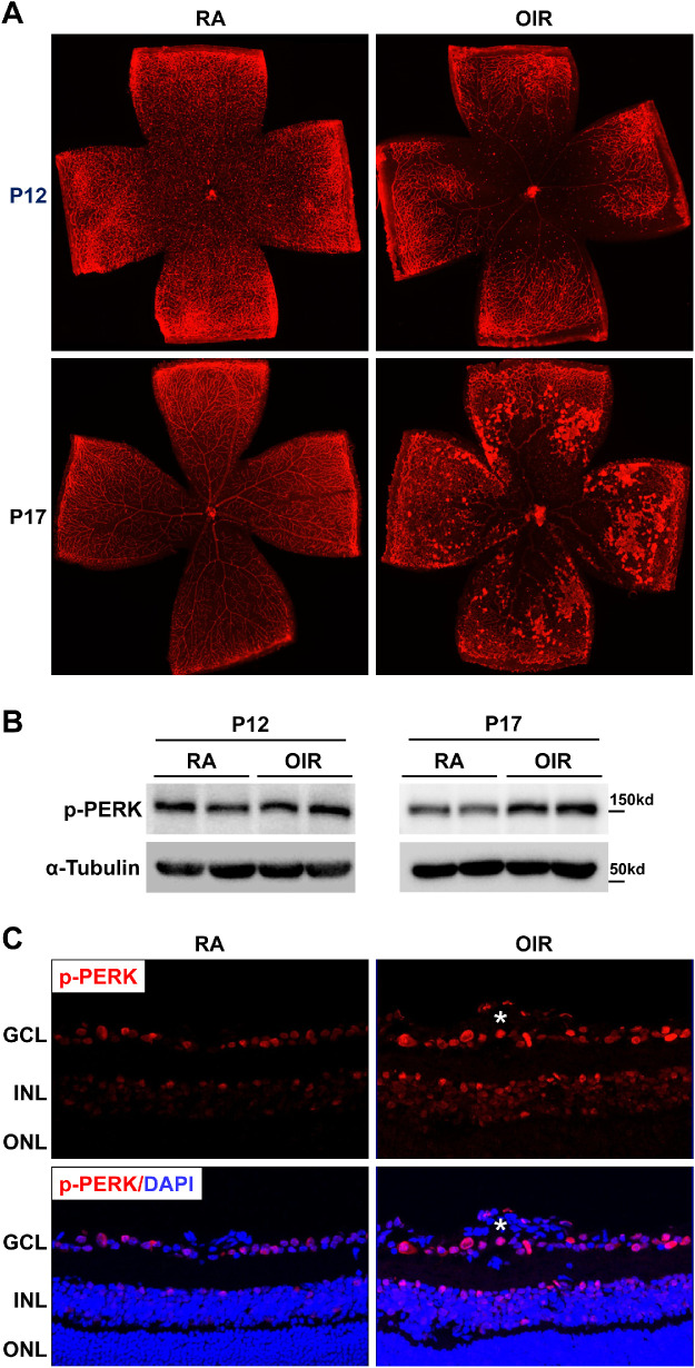
Purpose: Retinal ischemia is a common cause of a variety of eye diseases, such as retinopathy of prematurity, diabetic retinopathy, and vein occlusion. Protein kinase RNA-activated-like endoplasmic reticulum (ER) kinase (PERK), one of the main ER stress sensor proteins, has been involved in many diseases. In this study, we investigated the role of PERK in ischemia-induced retinopathy using a mouse model of oxygen-induced retinopathy (OIR).

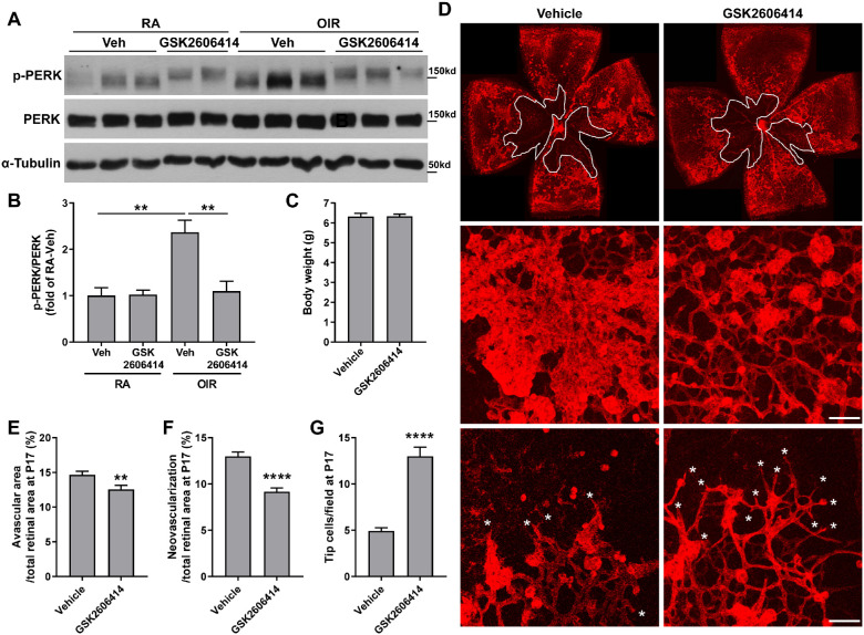
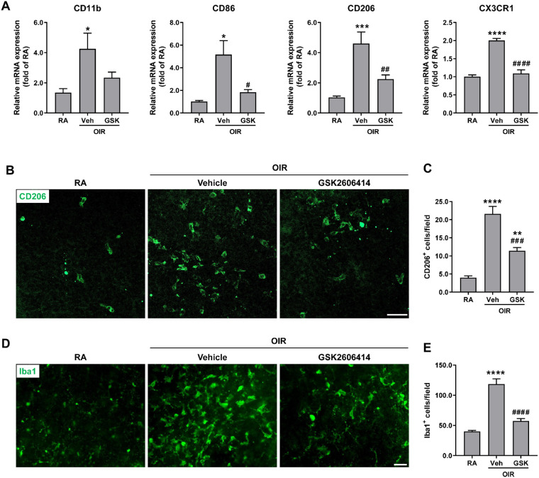
Methods: OIR was induced by subjecting neonatal pups to 70% oxygen at postnatal day 7 (P7) followed by returning to room air at P12. GSK2606414, a selective PERK inhibitor, was orally administrated to pups right after they were returned to room air once daily until 1 day before sample collection. Western blot, immunostaining, and quantitative PCR were used to assess PERK phosphorylation, retinal changes, and signaling pathways in relation to PERK inhibition.

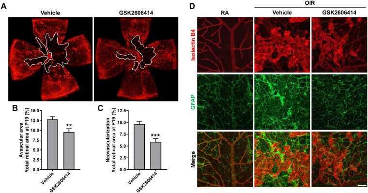
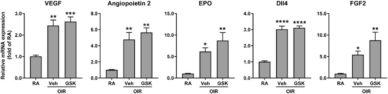
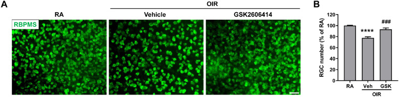
Results: PERK phosphorylation was prominently increased in OIR retinas, which was inhibited by GSK2606414. Concomitantly, PERK inhibition significantly reduced retinal neovascularization (NV) and retinal ganglion cell (RGC) loss, restored astrocyte network, and promoted revascularization. Furthermore, PERK inhibition downregulated the recruitment/proliferation of mononuclear phagocytes but did not affect OIR-upregulated canonical angiogenic pathways.

Conclusions: Our results demonstrate that PERK is involved in ischemia-induced retinopathy and its inhibition using GSK2606414 could offer an effective therapeutic intervention aimed at alleviating retinal NV while preventing neuron loss during retinal ischemia.


Conflict of interest statement

Disclosure: S. Shi, None; C. Ding, None; S. Zhu, None; F. Xia, None; S.E. Buscho, None; S. Li, None; M. Motamedi, None; H. Liu, None; W. Zhang, None

Figures

Figure 1.
Figure 1.
The phosphorylation level of PERK is increased in the retinas of OIR. WT mice were subjected to OIR or maintained in room air (RA) as control. Eyeballs or retinas were collected at P12 and P17. (A) Representative images of retinal flatmounts with Isolectin B4 staining for retinal vasculature at P12 and P17. (B) Phosphorylated PERK in the retina at P12 and P17 was evaluated by Western blot. The α-tubulin was used as the internal loading control. (C) Representative images of phosphorylated PERK in retinal sections at P17 were shown. DAPI (blue) was used to counterstain nuclei and identify the retinal nuclear layers as indicated by the labels. The white asterisk denotes the neovessel (n = 4). GCL, ganglion cell layer; INL, inner nuclear layer; ONL, outer nuclear layer.
Figure 2.
Figure 2.
PERK inhibition attenuates neovascularization and promotes revascularization in the retinas of OIR. Mice were subjected to OIR or maintained in RA as control, and they were treated with PERK inhibitor GSK2606414 (50 mg/kg) or vehicle from P12 to P16. Retinas or eyeballs were collected at P17. (A, B) Phosphorylated and total PERK was evaluated by Western blot. The α-tubulin was used as the internal loading control for normalization (n = 3-4; **P < 0.01). (C) Body weight of OIR mice treated with vehicle or GSK2606414 (n = 12). (D–G) Retinal flatmounts from vehicle- or GSK2606414-treated OIR mice at P17 were stained with Isolectin B4 and representative images were shown (upper panel). High magnification images were shown for neovascular tufts (middle panel) and tip cells (lower panel, white asterisks). Scale bar = 50 µm. Graphs represent the quantification of avascular and neovascularization area (n = 23–25) and the number of tip cells (n = 6). **P < 0.01 and ****P < 0.0001 compared with vehicle.
Figure 3.
Figure 3.
Blockade of PERK attenuates vasculopathy and preserves astrocyte network in OIR at P19. OIR mice were treated with GSK2606414 (50 mg/kg) or vehicle from P12 to P18. Eyeballs were collected at P19 and dissected for retinal flatmounts. (A–C) Retinal flatmounts were stained with Isolectin B4 for retinal vasculature. Graphs represent the quantification of avascular area and neovascularization area at P19 (n = 12). **P < 0.01 and ***P < 0.001 compared with vehicle. (D) Astrocytes were stained with anti-GFAP antibody (green) and retinal vessels were stained with Isolectin B4 (red). Scale bar = 50 µm.
Figure 4.
Figure 4.
Blockade of PERK does not affect canonical angiogenic pathway. OIR mice were treated with GSK2606414 (50 mg/kg) or vehicle from P12 to P16. Retinas were harvested at P17. The gene expressions of angiogenic factors were quantified by qPCR (n = 3-4). *P < 0.05, **P < 0.01, ***P < 0.001, and ****P < 0.0001 compared with RA controls.
Figure 5.
Figure 5.
Blockade of PERK attenuates the recruitment/proliferation of mononuclear phagocytes in OIR. The OIR mice were treated with GSK2606414 (50 mg/kg) or vehicle from P12 to P16. Retinas or eyeballs were harvested at P17. (A) Marker genes for different mononuclear phagocytes were quantified by qPCR (n = 3–7). (B–E) Representative images of CD206 and Iba1 staining in retinal flatmounts from vehicle- or GSK2606414-treated OIR mice at P17 were shown. Scale bar = 50 µm. Graphs represent the quantification of the number of CD206+ cells and Iba1+ cells (n = 5-6). *P < 0.05, **P < 0.01, ***P < 0.001, and ****P < 0.0001 compared with RA control; #P < 0.05, ##P < 0.01, ###P < 0.001, and ####P < 0.0001 compared with vehicle-treated OIR.
Figure 6.
Figure 6.
Blockade of PERK prevents RGC loss in OIR. The OIR mice were treated with GSK2606414 (50 mg/kg) or vehicle from P12 to P16. (A) Eyeballs were harvested at P17 and RGCs were stained with anti-RBPMS antibody (green) in retinal flatmounts. Scale bar = 50 µm. (B) Quantification of RGC number (n = 6). ****P < 0.0001 compared with RA control; ###P < 0.001 compared with vehicle-treated OIR.

References

    1. Campochiaro PA. Molecular pathogenesis of retinal and choroidal vascular diseases. Prog Retin Eye Res. 2015; 49: 67–81. - PMC - PubMed
    1. Kermorvant-Duchemin E, Sapieha P, Sirinyan M, et al. .. Understanding ischemic retinopathies: emerging concepts from oxygen-induced retinopathy. Doc Ophthalmol. 2010; 120(1): 51–60. - PubMed
    1. Fortmann SD, Grant MB.. Molecular mechanisms of retinal ischemia. Curr Opin Physiol. 2019; 7: 41–48. - PMC - PubMed
    1. Kim LA, D'Amore PA. A brief history of anti-VEGF for the treatment of ocular angiogenesis. Am J Pathol. 2012; 181(2): 376–379. - PMC - PubMed
    1. Stewart MW. The expanding role of vascular endothelial growth factor inhibitors in ophthalmology. Mayo Clin Proc. 2012; 87(1): 77–88. - PMC - PubMed

Publication types

MeSH terms