

Increased intracellular persulfide levels attenuate HlyU-mediated hemolysin transcriptional activation in Vibrio cholerae
- PMID: 37567478
- PMCID: PMC10509353
- DOI: 10.1016/j.jbc.2023.105147
Increased intracellular persulfide levels attenuate HlyU-mediated hemolysin transcriptional activation in Vibrio cholerae
Abstract
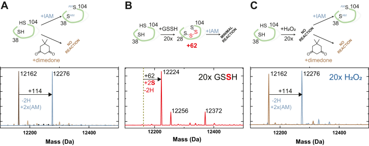
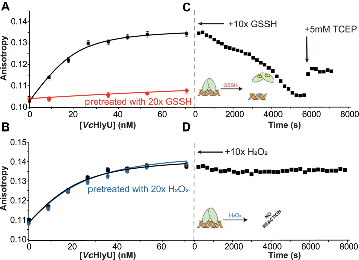
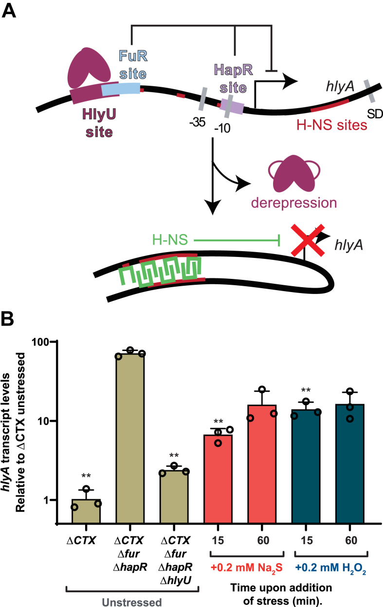
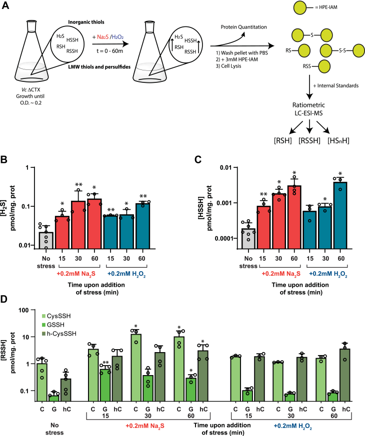
The vertebrate host's immune system and resident commensal bacteria deploy a range of highly reactive small molecules that provide a barrier against infections by microbial pathogens. Gut pathogens, such as Vibrio cholerae, sense and respond to these stressors by modulating the expression of exotoxins that are crucial for colonization. Here, we employ mass spectrometry-based profiling, metabolomics, expression assays, and biophysical approaches to show that transcriptional activation of the hemolysin gene hlyA in V. cholerae is regulated by intracellular forms of sulfur with sulfur-sulfur bonds, termed reactive sulfur species (RSS). We first present a comprehensive sequence similarity network analysis of the arsenic repressor superfamily of transcriptional regulators, where RSS and hydrogen peroxide sensors segregate into distinct clusters of sequences. We show that HlyU, transcriptional activator of hlyA in V. cholerae, belongs to the RSS-sensing cluster and readily reacts with organic persulfides, showing no reactivity or DNA dissociation following treatment with glutathione disulfide or hydrogen peroxide. Surprisingly, in V. cholerae cell cultures, both sulfide and peroxide treatment downregulate HlyU-dependent transcriptional activation of hlyA. However, RSS metabolite profiling shows that both sulfide and peroxide treatment raise the endogenous inorganic sulfide and disulfide levels to a similar extent, accounting for this crosstalk, and confirming that V. cholerae attenuates HlyU-mediated activation of hlyA in a specific response to intracellular RSS. These findings provide new evidence that gut pathogens may harness RSS-sensing as an evolutionary adaptation that allows them to overcome the gut inflammatory response by modulating the expression of exotoxins.
Keywords: Vibrio cholerae; bacterial pathogenesis; bacterial toxin; bacterial transcription; homocysteine; host–pathogen interaction; sulfur; thiol; transcription regulation.
Copyright © 2023 The Authors. Published by Elsevier Inc. All rights reserved.
Conflict of interest statement
Conflict of interest The authors declare that they have no conflicts of interest with the contents of this article.
Figures

Update of
-
Increased intracellular persulfide levels attenuate HlyU-mediated hemolysin transcriptional activation in Vibrio cholerae.bioRxiv [Preprint]. 2023 Mar 13:2023.03.13.532278. doi: 10.1101/2023.03.13.532278. bioRxiv. 2023. Update in: J Biol Chem. 2023 Sep;299(9):105147. doi: 10.1016/j.jbc.2023.105147. PMID: 36993174 Free PMC article. Updated. Preprint.
Similar articles
-
Sensing and regulation of reactive sulfur species (RSS) in bacteria.Curr Opin Chem Biol. 2023 Oct;76:102358. doi: 10.1016/j.cbpa.2023.102358. Epub 2023 Jul 1. Curr Opin Chem Biol. 2023. PMID: 37399745 Free PMC article. Review.
-
Increased intracellular persulfide levels attenuate HlyU-mediated hemolysin transcriptional activation in Vibrio cholerae.bioRxiv [Preprint]. 2023 Mar 13:2023.03.13.532278. doi: 10.1101/2023.03.13.532278. bioRxiv. 2023. Update in: J Biol Chem. 2023 Sep;299(9):105147. doi: 10.1016/j.jbc.2023.105147. PMID: 36993174 Free PMC article. Updated. Preprint.
-
Identification of the target DNA sequence and characterization of DNA binding features of HlyU, and suggestion of a redox switch for hlyA expression in the human pathogen Vibrio cholerae from in silico studies.Nucleic Acids Res. 2015 Feb 18;43(3):1407-17. doi: 10.1093/nar/gku1319. Epub 2015 Jan 20. Nucleic Acids Res. 2015. PMID: 25605793 Free PMC article.
-
Transcription of the Vibrio cholerae haemolysin gene, hlyA, and cloning of a positive regulatory locus, hlyU.Mol Microbiol. 1991 Aug;5(8):2031-8. doi: 10.1111/j.1365-2958.1991.tb00825.x. Mol Microbiol. 1991. PMID: 1766378
-
Spatiotemporal Regulation of Vibrio Exotoxins by HlyU and Other Transcriptional Regulators.Toxins (Basel). 2020 Aug 22;12(9):544. doi: 10.3390/toxins12090544. Toxins (Basel). 2020. PMID: 32842612 Free PMC article. Review.
Cited by
-
Sulfide-Responsive Transcription Control in Escherichia coli.Microorganisms. 2025 Feb 5;13(2):344. doi: 10.3390/microorganisms13020344. Microorganisms. 2025. PMID: 40005711 Free PMC article.
-
Sensing and regulation of reactive sulfur species (RSS) in bacteria.Curr Opin Chem Biol. 2023 Oct;76:102358. doi: 10.1016/j.cbpa.2023.102358. Epub 2023 Jul 1. Curr Opin Chem Biol. 2023. PMID: 37399745 Free PMC article. Review.
-
Bacterial Metallostasis: Metal Sensing, Metalloproteome Remodeling, and Metal Trafficking.Chem Rev. 2024 Dec 25;124(24):13574-13659. doi: 10.1021/acs.chemrev.4c00264. Epub 2024 Dec 10. Chem Rev. 2024. PMID: 39658019 Free PMC article. Review.
References
-
- Kathuria R., Chattopadhyay K. Vibrio cholerae cytolysin: multiple facets of the membrane interaction mechanism of a β-barrel pore-forming toxin. IUBMB Life. 2018;70:260–266. - PubMed
Publication types
MeSH terms
Substances
Grants and funding
LinkOut - more resources
Full Text Sources
