. 2023 Jul 27;12(15):4928.
doi: 10.3390/jcm12154928.

Paternal Age Matters: Association with Sperm Criteria's- Spermatozoa DNA Integrity and Methylation Profile

Affiliations

Paternal Age Matters: Association with Sperm Criteria's- Spermatozoa DNA Integrity and Methylation Profile

Marwa Lahimer et al. J Clin Med. .

Abstract

Advanced age has been reported to negatively affect sperm parameters and spermatozoa DNA integrity. A decline in sperm criteria was also associated with altered epigenetic marks such as DNA methylation with a potential downstream impact on in vitro fertilization success and clinical outcomes. The aim of the present retrospective study was to clarify the association between advanced paternal age (APA) and sperm parameters, DNA integrity and DNA methylation profile. A total of 671 patients consulting for infertility underwent sperm analysis, sperm DNA integrity assessment and methylation level measurement. The principal finding was that individuals over 40 years of age exhibit a significant increase in DNA fragmentation levels compared to the younger group (15% versus 9%, respectively, p = 0.04). However, there was no significant difference in DNA decondensation and sperm parameters in association with APA. In addition, a drop in the global methylation level was also found in men over 40 years (6% in the young group versus 2% in the old group, p = 0.03). As a conclusion, men over 40 years are at higher risk of elevated sperm DNA fragmentation and lower methylation level. Based on these observations, it is recommended that the assessment of sperm DNA fragmentation should be taken into consideration particularly after the age of 40. Our findings support the idea that paternal age is a crucial factor that should not be neglected during fertility evaluation and treatment since it is associated with epigenetics changes in sperm. Although the underlying mechanism remains to be clarified, we believe that environmental and professional exposure factors are likely involved in the process.

Keywords: paternal age; sperm parameters; spermatozoa DNA integrity and methylation.


Conflict of interest statement

The authors declare no conflict of interest.

Figures

Figure 1
Figure 1
Patients’ distribution according to conventional sperm parameters (The Sixth Edition of the WHO Manual for Human Semen Analysis 2021, 5th centile). Normospermic subjects: sperm concentration 16 × 106/mL; total motility ≥ 42%; typical forms ≥ 4%; viability ≥ 58%; volume ≥ 1.4 mL. Oligozoospermia (sperm concentration < 16 M/mL); Asthenozoospermia (% motility < 42%), Teratozoospermia (typical form < 4%); Oligoasthenoteratozoospermia (OATS) subjects: sperm concentration < 16 M/mL, total motility < 42%, typical form < 4%); reduced volume (hypo-spermia) < 1.4 mL). Increased volume (hyper-spermia) (95% confidence) volume > 6.2 mL). *: Oligoasthenoteratozoospermia [35].
Figure 2
Figure 2
Impact of advanced paternal age on sperm DNA integrity. (A) DFI was relatively high in G1 compared to G2, * p < 0.05, no significant difference in SDI between the two groups. DNA fragmentation refers to the physical breakage or damage to the DNA strands within sperm cells. (B) Detection of single- or double-strand breaks in sperm DNA. The fragmented sperm DNA is visualized as a green color conversely, non-fragmented sperm DNA is represented by a blue color using the DAPI dye. (C) Microscopic observation of the aniline blue staining, the blue color indicates the penetration of aniline blue into decondensed chromatin within the spermatozoa. It represents an indicator of the spermatozoa’s maturity.
Figure 3
Figure 3
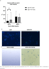
Impact of advanced paternal age on DNA methylation profile. (A) Decrease in sperm DNA methylation levels in G2 compared to G1, * p < 0.05. (B) Detection of 5′-methylcytosine in spermatozoa, the binding of the anti-5-methylcytosine antibody is denoted by the green color, while the absence of binding is visualized as a blue color using the DAPI dye.
Figure 3
Figure 3
Impact of advanced paternal age on DNA methylation profile. (A) Decrease in sperm DNA methylation levels in G2 compared to G1, * p < 0.05. (B) Detection of 5′-methylcytosine in spermatozoa, the binding of the anti-5-methylcytosine antibody is denoted by the green color, while the absence of binding is visualized as a blue color using the DAPI dye.

References

    1. Huang C., Li B., Xu K., Liu D., Hu J., Yang Y., Nie H., Fan L., Zhu W. Decline in semen quality among 30,636 young Chinese men from 2001 to 2015. Fertil. Steril. 2017;107:83–88.e2. doi: 10.1016/j.fertnstert.2016.09.035. - DOI - PubMed
    1. Barsky M., Blesson C.S. Should we be worried about advanced paternal age? Fertil. Steril. 2020;114:259–260. doi: 10.1016/j.fertnstert.2020.04.033. - DOI - PubMed
    1. Bahri H., Ben Khalifa M., Ben Rhouma M., Abidi Z., Abbassi E., Ben Rhouma K., Benkhalifa M. Decline in semen quality of North Afri-can men: A retrospective study of 20,958 sperm analyses of men from different North African countries tested in Tunisia over a period of 6 years (2013–2018) Ann. Hum. Biol. 2021;48:350–359. doi: 10.1080/03014460.2021.1957501. - DOI - PubMed
    1. Jimbo M., Kunisaki J., Ghaed M., Yu V., Flores H.A., Hotaling J.M. Fertility in the aging male: A systematic review. Fertil. Steril. 2022;118:1022–1034. doi: 10.1016/j.fertnstert.2022.10.035. - DOI - PMC - PubMed
    1. Kaarouch I., Bouamoud N., Madkour A., Louanjli N., Saadani B., Assou S., Aboulmaouahib S., Amzazi S., Copin H., Benkhalifa M., et al. Paternal age: Negative impact on sperm genome decays and IVF outcomes after 40 years. Mol. Reprod. Dev. 2018;85:271–280. doi: 10.1002/mrd.22963. - DOI - PubMed