. 2023 Jul 29;12(15):5002.
doi: 10.3390/jcm12155002.

Computational and Experimental Analyses for Pathogenicity Prediction of ACVRL1 Missense Variants in Hereditary Hemorrhagic Telangiectasia

Affiliations

Computational and Experimental Analyses for Pathogenicity Prediction of ACVRL1 Missense Variants in Hereditary Hemorrhagic Telangiectasia

Toru Iwasa et al. J Clin Med. .

Abstract

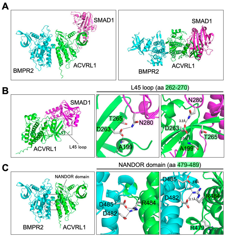
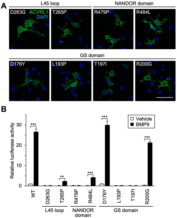
Hereditary hemorrhagic telangiectasia (HHT) is a vascular disease caused by the defects of ALK1/ACVRL1 receptor signaling. In this study, we evaluated 25 recently identified ACVRL1 missense variants using multiple computational pathogenicity classifiers and experimentally characterized their signal transduction capacity. Three extracellular residue variants showed no detectable cell surface expression and impairment of bone morphogenetic protein 9 (BMP9) responsiveness of SMAD-dependent transcription in luciferase assays. Four variants with amino acid replacement in the motifs essential for the intracellular kinase function lost SMAD-dependent signaling. Most of other variations in the kinase domain also caused marked downregulation of signaling; however, two variants behaved as the wild-type ACVRL1 did, while computational classifiers predicted their functional abnormalities. Three-dimensional structure prediction using the ColabFold program supported the significance of the L45 loop and NANDOR domain of ACVRL1 for its association with SMAD1 and BMPR2, respectively, and the variations in these motifs resulted in the reduction of SMAD signaling. On the other hand, two of the GS domain variants maintained high signal transduction capacity, which did not accord with their computational pathogenicity prediction. These results affirm the requirement of a combinatory approach using computational and experimental analyses to accurately predict the pathogenicity of ACVRL1 missense variants in the HHT patients.

Keywords: ACVRL1; ALK1; BMP; SMAD signaling; hereditary hemorrhagic telangiectasia.


Conflict of interest statement

The authors declare no conflict of interest.

Figures

Figure 1
Figure 1
Characteristics of ACVRL1 missense variants selected for computational and experimental analyses. In the schematic structure of ACVRL1, the functional motifs/domains conserved among ALK family receptors are filled in grey, and those shared by protein kinases are filled in black. AxK, AxK motif; DLG, DLG motif; GS, GS domain; HRD, HRD domain; L45, L45 loop; Likely Patho, Likely pathogenic; NANDOR, NANDOR domain; n.d., not deposited; TM, transmembrane domain; Uncertain, Uncertain significance; VUS, variant of uncertain significance. References: V32E [19,20], T52P [21], C90W [20], D176Y [24], L193P [22], T197I [25], R200G [19,20], V205G [19,20], K229R [26], D263G [37], T265P [24], H280R [19,20,28,30], L300P [20], H328Y [19,20,27,29], D330Y [10,31,32], D348E [20], P424T [38], P433S [33,34], D437G [35], C443R [20], P449A [20], C471Y [20], R479P [28], R484L [36].
Figure 2
Figure 2
Subcellular localization and signal transduction capacity of extracellular residue variants. (A) The extracellular residue variants were not detected on the cell surface. Immunocytochemistry in non-permeabilized NIH-3T3 cells. Scale bar: 40 μm. (B) The extracellular residue variants were detected in the cytoplasm of permeabilized NIH-3T3 cells. Scale bar: 40 μm. (C) The extracellular residue variants showed marked down regulation of BMP9-induced signal transduction in BRE-luciferase assays. Results are expressed as fold induction over the value obtained for the cells expressing wild-type (WT) ACVRL1 with the vehicle treatment. *** p < 0.001 (Tukey’s test).
Figure 3
Figure 3
Subcellular localization and signal transduction capacity of kinase domain variants. (A) All kinase domain variants were detected on the cell surface. Immunocytochemistry in non-permeabilized NIH-3T3 cells. Scale bar: 40 μm. (B) Most of the kinase domain variants failed to mediate BMP9-induced transcriptional activation, while the D437G variant showed weak but statistically significant signal transduction in BRE-luciferase assays. In contrast, V205G and D235Y variants kept high BMP9-induced signaling activity comparable to that of wild-type ACVRL1. Results are expressed as fold induction over the value obtained for the cells expressing wild-type (WT) ACVRL1 with the vehicle treatment. The data for wild-type ACVRL1 are same as those in Figure 2C. *** p < 0.001 (Tukey’s test).
Figure 4
Figure 4
Three-dimensional structural prediction of ACVRL1 interaction with SMAD1 and BMPR2. ACVRL1 kinase domain (green; aa 195-503), BMPR2 kinase domain (cyan; aa 197-512) and SMAD1 MH2 domain (magenta; aa 271-465) were applied to the structural prediction using ColabFold. (A) The predicted structure of the ACVRL1-BMPR2-SMAD1 trimer viewed from two different directions. A top view of the left image is on the right. (B) ACVRL1-SMAD1 dimer structure (left) with an enlarged view of the dotted box (middle). The figure on the right shows the area in the middle figure viewed from another direction. Side chains of ACVRL1 D263 and T265, that of SMAD1 N280 and a main chain of ACVRL1 A199 are depicted. In the ACVRL1-SMAD1 interface shown on the right, the interaction between ACVRL1 D263 in the L45 loop and SMAD1 N280 was predicted. Intramolecular interaction between ACVRL1 D263 and A199 was also indicated. D263 and T265 of ACVRL1 are localized in the same β sheet. (C) ACVRL1-BMPR2 dimer structure (left) with an enlarged view of the dotted box (middle). The figure on the right depicts the image in the middle figure viewed from another direction. In the ACVRL1-BMPR2 interface shown on the right, the interaction between ACVRL1 R484 in the NANDOR domain with BMPR2 D482 and D485 was predicted. On the other hand, ACVRL1 R479 did not show direct interaction with BMPR2. Carbon, oxygen and nitrogen atoms are represented by gray, red and blue, respectively, and hydrogen atoms are omitted for clarity. Dotted lines indicate possible amino acid interaction with the distance less than 4 Å.
Figure 5
Figure 5
Influence of missense variations in functional domains conserved in ALK family receptors. (A) ACVRL1 receptors with a variation in the L45 loop, NANDOR domain or GS domain were localized on the cell surface. Immunocytochemistry in non-permeabilized NIH-3T3 cells. Scale bar: 40 μm. (B) Missense variations in those functional domains markedly down regulated BMP9-induced transcription in BRE-luciferase assays while that mediated by T265P and R484L variants still had statistical significance. In contrast, the cells expressing D176Y and R200G variants showed high transcriptional activation in response to BMP9. Results are expressed as fold induction over the value obtained for the cells expressing wild-type (WT) ACVRL1 with the vehicle treatment. The data for wild-type ACVRL1 are same as those in Figure 2C. ** p < 0.01; *** p < 0.001 (Tukey’s test).
Figure 6
Figure 6
Signal transduction capacity of D176Y, R200G, V205G and D235Y variants. (A) D176Y, R200G, V205G and D235Y variants showed BMP9 dose dependency similar to wild-type (WT) ACVRL1 in BRE-luciferase assays. Results are expressed as fold induction over the value obtained for the cells expressing wild-type ACVRL1 with the vehicle treatment. * p < 0.05; ** p < 0.01; *** p < 0.001 (Dunnett’s test). (B,C) The cells expressing D176Y, R200G, V205G and D235Y variants showed BMP9-induced transcriptional activity in ID1- or BMPR2-luciferase assays. Results are expressed as fold induction over the values obtained for vehicle-treated wild-type ACVRL1. *** p < 0.001 (Tukey’s test). (D) The cells expressing D176Y, R200G, V205G and D235Y variants showed SMAD1/5/9 phosphorylation in response to the BMP9 treatment, which was comparable to those expressing wild-type ACVRL1. Note that vector-transfected cells did not show significant SMAD1/5/9 phosphorylation. Western blot analysis.
Figure 7
Figure 7
Responsiveness of wild-type ACVRL1 and missense variants to endoglin co-expression. (A) Transcription mediated by wild-type (WT) ACVRL1 was increased by the co-expression of endoglin (ENG) in BRE-luciferase assays. Results are expressed as fold induction over the value obtained for the cells with wild-type ACVRL1 expression and vehicle treatment. ** p < 0.01; *** p < 0.001 (Tukey’s test). (B) D176Y, R200G, V205G and D235Y variants also showed significant induction of BMP9-dependent signal transduction when co-expressed with endoglin. BRE-luciferase assays. *** p < 0.001 (Tukey’s test). (C) Co-expression of endoglin significantly increased the BMP9-induced transcription mediated by K229R, T265P and D348E. In contrast, endoglin significantly decreased the BMP9-induced transcription in the cells expressing R484L. BRE-luciferase assays. In panels B and C, the results are expressed as fold induction over the value obtained for the cells with wild-type ACVRL1 expression and vehicle treatment. The results in panel A serve as a wild-type ACVRL1 control from the same experiment for panel (C). * p < 0.05; *** p < 0.001 (Tukey’s test).

Similar articles

References

    1. Shovlin C.L., Guttmacher A.E., Buscarini E., Faughnan M.E., Hyland R.H., Westermann C.J., Kjeldsen A.D., Plauchu H. Diagnostic criteria for hereditary hemorrhagic telangiectasia (Rendu-Osler-Weber syndrome) Am. J. Med. Genet. 2000;91:66–67. doi: 10.1002/(SICI)1096-8628(20000306)91:1<66::AID-AJMG12>3.0.CO;2-P. - DOI - PubMed
    1. Bernabeu C., Bayrak-Toydemir P., McDonald J., Letarte M. Potential Second-Hits in Hereditary Hemorrhagic Telangiectasia. J. Clin. Med. 2020;9:3571. doi: 10.3390/jcm9113571. - DOI - PMC - PubMed
    1. Robert F., Desroches-Castan A., Bailly S., Dupuis-Girod S., Feige J.J. Future treatments for hereditary hemorrhagic telangiectasia. Orphanet J. Rare Dis. 2020;15:4. doi: 10.1186/s13023-019-1281-4. - DOI - PMC - PubMed
    1. Oh S.P., Seki T., Goss K.A., Imamura T., Yi Y., Donahoe P.K., Li L., Miyazono K., ten Dijke P., Kim S., et al. Activin receptor-like kinase 1 modulates transforming growth factor-beta 1 signaling in the regulation of angiogenesis. Proc. Natl. Acad. Sci. USA. 2000;97:2626–2631. doi: 10.1073/pnas.97.6.2626. - DOI - PMC - PubMed
    1. Urness L.D., Sorensen L.K., Li D.Y. Arteriovenous malformations in mice lacking activin receptor-like kinase-1. Nat. Genet. 2000;26:328–331. doi: 10.1038/81634. - DOI - PubMed

LinkOut - more resources