Case Reports
. 2023 Jul 30;12(15):5009.
doi: 10.3390/jcm12155009.

Nerve MR in the Differential Diagnosis of Neuropathies: A Case Series from a Single Center

Affiliations
Case Reports

Nerve MR in the Differential Diagnosis of Neuropathies: A Case Series from a Single Center

Carolina Giordano et al. J Clin Med. .

Abstract

In the present study, through a case series, we highlighted the role of magnetic resonance (MR) in the identification and diagnosis of peripheral neuropathies. MR neurography allows the evaluation of the course of nerves through 2D and 3D STIR sequences with an isotropic voxel, whereas the relationship between nerves, vessels, osteo-ligamentous and muscular structures can be appraised with T1 sequences. Currently, DTI and tractography are mainly used for experimental purposes. MR neurography can be useful in detecting subtle nerve alterations, even before the onset of symptoms. However, despite being sensitive, MR neurography is not specific in detecting nerve injury and requires careful interpretation. For this reason, MR information should always be supported by instrumental clinical tests.

Keywords: magnetic resonance neurography; peripheral nervous system; peripheral neuropathy.


Conflict of interest statement

The authors declare no conflict of interest.

Figures

Figure 1
Figure 1
MR T2-weighted (top) and T1-weighted fat post-contrast contrast images (bottom). The images in (A) are in the coronal plane, those in (B) are in the sagittal plane and those in (C) are in the axial plane. The images show thickened and pathological cranial nerves with a cystic appearance, high signals in T2w images and almost no contrast enhancement. In both (A), yellow arrows indicate pathological V3 branches of the trigeminal nerve (transverse diameter after the foramen ovale: left 3.8 mm; right 10 mm); in both (B), yellow arrows indicate pathological facial nerve in the intraparotid course (transverse diameter in intraparotid course: left 6.7 mm vs. right 2.2 mm); in both (C), yellow arrows indicate pathological right V1 branch of the trigeminal nerve (transverse diameter after superior orbital foramen: right 6.9 mm vs. left 2.3 mm).
Figure 2
Figure 2
MR (multiplanar reconstructions) of 3D cube neurography images show pathological, thickened cranial nerves along their extracranial course, with some cystic-like components. The images show the involvement of trigeminal branches V1 in ((D)—yellow star), V2 in ((B,D)—curved arrows with a transverse diameter after inferior orbital foramen: left 1.8 mm; right 2.6 mm) and V3 from its passage in the foramen ovale (asterisks in (A,B)) to its lingual (arrowheads in (B,D)) and alveolar divisions (thick arrows in (BD)); the involvement of the left facial nerve in its intraparotid course (thin arrows in (A,C)) and the hypoglossal nerve (open arrow in (B), transverse diameter at C1 level: left 4.6 mm; right 2.1 mm).
Figure 3
Figure 3
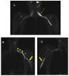
MIP (Maximum Intensity Projection) reconstructions of the 3D cube nerve MR sequence of the brachial (A) and lumbosacral (B) plexus showing a pathological and asymmetric thickening and increase in T2 signal representation of them.
Figure 4
Figure 4
MR 3D cube nerve images show thickening and T2 hyperintensity of the left obturator nerve (thin arrow in A) and lumbosacral trunk (thick arrow in B) compared to the contralateral one (transverse diameter on left L5 root 7.7 mm (n.v. * 5.99 ± 0.66) and on left S1 root 9.1 mm (n.v. * 5.27 ± 0.53). * Normal values (n.v.) according to the recent study of Su X et al. [20].
Figure 5
Figure 5
MR 3D cube nerve images show thickening and T2 hyperintensity of brachial plexus and in particular in (A), we can observe a pathologic left C6 root (thin arrow transverse diameter of C6 left root 10.3 mm; n.v. * 4.57 ± 0.51); in B, we can observe the involvement of left trunks and cords (arrowheads) and in (B,C), we can observe the involvement of the peripheral nerves of the bilateral arms (thick arrows). * Normal values (n.v.) according to the recent study of Su X et al. [20].
Figure 6
Figure 6
MIP (Maximum Intensity Projection) reconstructions of the 3D cube nerve MR sequence of the lumbosacral plexus show regular size and signal intensity of lumbar and sacral roots.
Figure 7
Figure 7
Axial 2D MR of sciatic nerve; on the left are the T1w images and on the right are the T2 STIR images. Bilateral and symmetrical sciatic nerve T2 enlargement and fascicular hyperintensity at the level of the gluteal region (thin arrows (B); CSA of sciatic nerve of 103 mm2 on the right; 90.1 mm2 on the left) and at the mid-thigh (thin arrows in (D,F)). Mixed, acute and chronic denervation of the muscles of the anterolateral compartment of the thigh with denervation edema (hyperintensity in STIR, arrowheads in (D,F)) and fat replacement (thick arrows in (C,E)). Mainly chronic denervation of the gluteal muscles (hyperintensity in T1w, thick arrows in (A)).
Figure 8
Figure 8
MIP (Maximum Intensity Projection) reconstructions of the 3D cube nerve-MR sequence of the lumbosacral plexus documented a moderate and diffuse increase in thickness and signal intensity on T2W images of the lumbar and sacral roots; the left S1root (arrows) appears to be pathological (thickened and markedly hyperintense in T2w). Transverse diameter S1 roots (n.v. * 5.27 ± 0.53) left 8.4 mm; right 8.2 mm). * Normal values (n.v.) according to the recent study of Su X et al. [20].
Figure 9
Figure 9
Pathological alterations of the left sciatic nerve (thin arrows in (B,D)), characterized by a significant increase in volume (CSA in the upper third of the thigh of about 123 mm2) with associated fascicular hypertrophy and an increase in signals in T2w images, especially in the height of the middle and lower third of the thigh and with extension to the tibial nerve (thin arrow in (F)), up to the lower third of the leg. Also, in the right sciatic nerve, there are some alterations (thin arrows in (B,D), much more tenuous than the contralateral nerve, characterized by an increase in the thickness of the nerve (CSA at the upper third of the thigh of about 110 mm2), a moderate increase signal in T2w images and fascicular hypertrophy, without evident tibial nerve signal alterations. Mixed denervation, acute and chronic, of left and right semimembranosus and posteromedial compartment muscles of the left leg (muscle hyperintensity and fascial edema in STIR, thick arrows in (D,F)) and fat replacement (muscle bundle atrophy with replacement of hyperintense tissue at T1W in (A,C,E), more evident in some areas: arrowheads in (C,E)).

References

    1. Martyn C.N., Hughes R.A. Epidemiology of peripheral neuropathy. J. Neurol. Neurosurg. Psychiatry. 1997;62:310–318. doi: 10.1136/jnnp.62.4.310. - DOI - PMC - PubMed
    1. Iqbal Z., Azmi S., Yadav R., Ferdousi M., Kumar M., Cuthbertson D.J., Lim J., Malik R.A., Alam U. Diabetic Peripheral Neuropathy: Epidemiology, Diagnosis, and Pharmacotherapy. Clin. Ther. 2018;40:828–849. doi: 10.1016/j.clinthera.2018.04.001. - DOI - PubMed
    1. Ricci L., Luigetti M., Florio L., Capone F., Di Lazzaro V. Causes of chronic neuropathies: A single-center experience. Neurol. Sci. 2019;40:1611–1617. doi: 10.1007/s10072-019-03899-z. - DOI - PubMed
    1. England J.D., Asbury A.K. Peripheral neuropathy. Lancet. 2004;363:2151–2161. doi: 10.1016/S0140-6736(04)16508-2. - DOI - PubMed
    1. Boerkoel C.F., Takashima H., Garcia C.A., Olney R.K., Johnson J., Berry K., Russo P., Kennedy S., Teebi A.S., Scavina M., et al. Charcot-Marie-Tooth disease and related neuropathies: Mutation distribution and genotype-phenotype correlation. Ann. Neurol. 2001;51:190–201. doi: 10.1002/ana.10089. - DOI - PubMed

Publication types

LinkOut - more resources