. 2023 Jul 25;15(15):3758.
doi: 10.3390/cancers15153758.

Radical Tumor Denervation Activates Potent Local and Global Cancer Treatment

Affiliations

Radical Tumor Denervation Activates Potent Local and Global Cancer Treatment

John D Mitsou et al. Cancers (Basel). .

Abstract

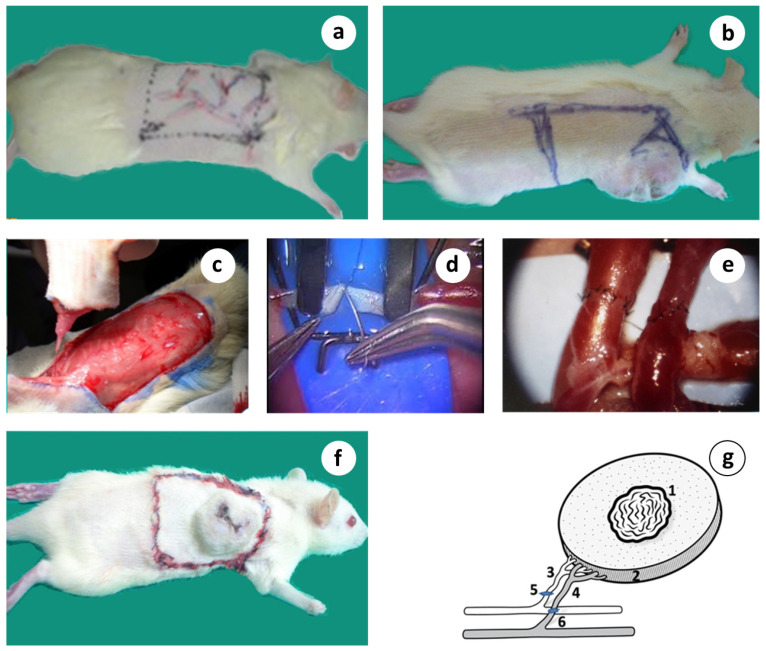
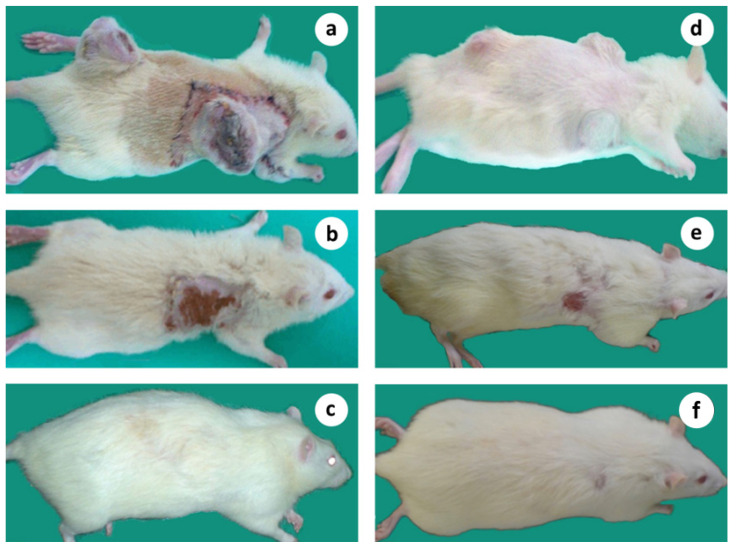
This preliminary study seeks to determine the effect of R&P denervation on tumor growth and survival in immunocompetent rats bearing an aggressive and metastatic breast solid tumor. A novel microsurgical approach was applied "in situ", aiming to induce R&P denervation through the division of every single nerve fiber connecting the host with the primary tumor via its complete detachment and re-attachment, by resecting and reconnecting its supplying artery and vein (anastomosis). This preparation, known as microsurgical graft or flap, is radically denervated by definition, but also effectively delays or even impedes the return of innervation for a significant period of time, thus creating a critical and therapeutic time window. Mammary adenocarcinoma cells (HH-16.cl4) were injected into immunocompetent Sprague Dawley adult rats. When the tumors reached a certain volume, the subjects entered the study. The primary tumor, including a substantial amount of peritumoral tissue, was surgically isolated on a dominant artery and vein, which was resected and reconnected using a surgical microscope (orthotopic tumor auto-transplantation). Intending to simulate metastasis, two or three tumors were simultaneously implanted and only one was treated, using the surgical technique described herein. Primary tumor regression was observed in all of the microsurgically treated subjects, associated with a potent systemic anticancer effect and prolonged survival. In stark contrast, the subjects received a close to identical surgical operation; however, with the intact neurovascular connection, they did not achieve the therapeutic result. Animals bearing multiple tumors and receiving the same treatment in only one tumor exhibited regression in both the "primary" and remote- untreated tumors at a clinically significant percentage, with regression occurring in more than half of the treated subjects. A novel therapeutic approach is presented, which induces the permanent regression of primary and, notably, remote tumors, as well as, evidently, the naturally occurring metastatic lesions, at a high rate. This strategy is aligned with the impetus that comes from the current translational research data, focusing on the abrogation of the neuro-tumoral interaction as an alternative treatment strategy. More data regarding the clinical significance of this are expected to come up from a pilot clinical trial that is ongoing.

Keywords: abscopal effect; cancer neurobiology; microsurgery; radical denervation; tumor denervation; tumor regression.


Conflict of interest statement

There are no financial conflict of interest to disclose.

Figures

Figure 1
Figure 1
Microscopic histopathological evidence of malignancy and macroscopically visible lung metastases. (a) Primary tumor; (b) Lung metastasis; (c,d) Macroscopically visible lung metastases.
Figure 2
Figure 2
Micro PET scanning demonstration of primary and metastatic lesions in 3 different subjects. (a) There is a large tumor located in the right thoracic wall posteriorly demonstrating heterogeneous tracer distribution (indicative SUVmax: 3.9) and photopenic areas indicative of necrosis. Also, there is a second soft tissue mass in the right lateral chest wall close to the aforementioned large tumor likely representing a regional lymph node (SUVmax 4.2). In addition, there are multiple metastatic lung lesions bilaterally (indicative SUVmax 3.9), (b) There is a large tumor located in the right thoracic wall posteriorly demonstrating heterogeneous tracer distribution (indicative SUVmax: 11.2) and photopenic areas indicative of necrosis. In addition, there is a second soft tissue mass in the right lateral chest wall close to the aforementioned large tumor likely representing a regional lymph node (SUVmax 9.7). Focal FDG uptake (SUVmax 2.4) is also noted in what appears to be an additional lymph node in the left side of the thoracic cage superiorly, (c) There is a large tumor located in the right thoracic wall posteriorly demonstrating heterogeneous tracer distribution (indicative SUVmax: 8.8) and photopenic areas indicative of necrosis. Also, there is a second soft tissue mass in the right anterolateral chest wall close to the aforementioned large tumor likely representing a regional lymph node (SUVmax 10.0).
Figure 3
Figure 3
A step-by-step description of the microsurgical orthotopic tumor auto-transplantation. (a) Design of the proximal dorsolateral adipo-myocutaneous flap with its vascular tree marked; (b) Tumor about 6 wk. after induction with the tumor complex marked; (c) Surgical preparation of the vascular tumor complex graft isolated on its dominant vascular pedicle; (d) Standard hand-sewn microvascular anastomosis; (e) Microvascular anastomosis of both vessels completed; (f) Microsurgical tumor complex in place consisted by the primary tumor including a substantial amount of healthy tissue; (g) Schematic presentation of the microsurgical tumor composite graft based on its artery and vein; 1 = tumor, 2 = peritumoral healthy area, 3 = circumflex scapular artery, 4 = circumflex scapular vein, 5 and 6 = microvascular anastomoses.
Figure 4
Figure 4
Adverse wound healing effects in microsurgical flaps and contrasting differences with the intact vascular pedicle flap, in rabbits although these 2 surgical preparations are similar regarding the blood flow dynamics. (a) The original size of the microsurgical and the vascular pedicle flap (day 0); (b) Severe contraction, trophic ulcers, and abortive hair growth inside the microsurgical flap area (day 30); (c) In strike contrast, vascular pedicle flap shows superabundant hair growth, light contraction without trophic ulcers (day 30); (d) The real size of the vascular pedicle flap after hair removal showing less contraction and comparing; (e) No evidence of revascularization from the underline vascular bed in a microsurgical flap which is easily separated without dissection or bleeding (day 30), (unpublished data).
Figure 5
Figure 5
(a) Vascular pedicle or Vascular tumor complex; (1) Tumor; (2) Peritumoral healthy tissue and the underlying muscle; (3) Circumflex subscapular artery; (4) Circumflex subscapular vein. (b) Neurovascular pedicle or neurovascular tumor complex; (1) Tumor; (2) Peritumoral healthy tissue and the underlying muscle; (3) Circumflex subscapular artery; (4) Circumflex subscapular vein; (5) Accompanying nerve.
Figure 6
Figure 6
(a) Collective, comparative results on primary tumor volume changes. Redline, no treatment normal control (group 1); An almost linear, and sharp increase in the tumor size. Blueline, partial denervation, neurovascular groups with or without membrane (group 4 and 7): A similar type but with a moderate increase was shown. Greenline, radical and persistent (R&P) denervation, microsurgical groups with or without membrane (group 2 and 6); After an initial, light increase during the first 2 wk. after treatment, tumor deteriorates gradually until its complete and irreversible regression in microsurgical group 2 (*: p value < 0.005 in comparison of microsurgical tumor complex group with both control and neurovascular tumor complex groups). (b) Analytical presentation of the primary tumor volume changes in microsurgical group 2; after an initial moderate growth for about 2 wk. the tumors started to constantly decrease in size until their elimination, in about 5–7 wk. after treatment. (c) Collective results on LTS rate: from left to right: 0% = Normal control group, no treatment (group 1); 87.5% = microsurgical groups, R&P denervation (groups 2 and 6), 12.5% = neurovascular groups, partial denervation (groups 4 and 7); 57.1% = microsurgical group with 2 or 3 tumors (group 10); 0% = 2-step destruction of tissue continuity, with or without membrane (groups 5 and 8).
Figure 7
Figure 7
Gradual shrinkage of the transplanted tumor, followed by complete tumor regression in about 6 wk. after treatment, leading to relapse-free survival for more than 12 months (follow up period). (a) Microsurgical tumor complex 2 wk. after R&P denervation; (bd) Steady reduction in the size of the primary tumor; (e,f). Primary tumor regressed following a classical wound-healing contraction pattern.
Figure 8
Figure 8
Animals survived more than one year after microsurgical tumor auto-transplantation. (a) 13 months; (b) 15 months; (c) 18 months after treatment.
Figure 9
Figure 9
Comparison of LTS rate (a) and MS time (b) vs. the classified relative estimation of the reinnervation potential uncovered an inverse relationship of the therapeutic response to the reinnervation potential. Minimum (group 2) = microsurgical group with membrane; Close to minimum (group 6) = microsurgical group without membrane; Moderate (group 3) = vascular group; Close to optimum (group 4) = neurovascular group with membrane; Optimum (group 7) = neurovascular group without membrane. Overall LTS rate (c) and MS time (d) vs. microvascular anastomosis performance; Groups 2, 6 = microsurgical tumor complex with or without membrane, Groups 1, 3, 4, 7, 9 = Normal control, Vascular group, Neurovascular group with or without membrane, and Ischemia sham control group.
Figure 10
Figure 10
(a) Cumulative results of the MS time in all operated but non-cured subjects has shown that it is significantly less (p < 0.05) compared to the untreated, normal control animals; (b) LTS rate and MS time (c) correlation after destruction of host–tumor complex tissue continuity: Destruction of host-tumor continuity (groups 2, 6) = microsurgical group with or without membrane, Close to total destruction, no nerve (group 3) = vascular group, Close to total destruction, with nerve (groups 4, 7, 9) = neurovascular group with or without membrane, and no destruction at all (group 1) = Normal control group. LTS rate (d) MS time and (e) correlation after the destruction of host–tumor complex tissue continuity in 1 versus 2 surgical steps shows that destruction in 2 steps eliminates the possibility for long-term survival, significantly suppresses the MST time: Destruction of tissue continuity in 1 step = microsurgical group with or without membrane (groups 2, 6), destruction in 2 steps, with or without membrane (groups 5, 6). Comparison of survival of the microsurgical (f,g) and neurovascular groups (h,i) in correlation to membrane insertion, shows no significant differences in both, LTS rate and MS time: Microsurgical group with or without membrane (groups 2, 6), Neurovascular group with or without membrane (groups 4, 7).
Figure 11
Figure 11
LTS rate (a) and MS time (b) vs. surgical trauma severity. Animal groups with identical or close to identical trauma have shown wide variations on both parameters, indicating no direct influence of trauma severity on the results: Microsurgical tumor complex with membrane (group 2), Vascular group without membrane (group 4), Neurovascular group with membrane (group 4). Microsurgical tumor complex without membrane (group 6), Neurovascular group without membrane (group 7), Tumor ischemia sham-control (group 9). LTS rate (c) and MS time (d) vs. classified, relative estimation. A proportional relationship of the therapeutic response to the degree of the promptly induced surgical denervation has been recorded. R&P denervation (groups 2, 6) = Microsurgical tumor complex with or without membrane; Partial but severe denervation (group 3) = vascular tumor complex, Partial moderate denervation (groups 4, 7) = neurovascular tumor complex with or without membrane; No denervation (group 1) = Normal control.
Figure 12
Figure 12
Microsurgical tumor complex in a subject bearing 2, simultaneously induced tumors; (a) Animal with 2 tumors 7–8 wk. after tumor induction, on the day of treatment, applied to only one of the tumors; (b) The same animal showing the regression of both tumors, 10 wk. after treatment; (c) The cured animal 14 months after tumor induction without relapse of malignancy. (df) Microsurgical tumor complex in a subject bearing 3, simultaneously induced tumors; (d) Animal with 3 tumors, 5 wk. after tumor induction; (e) The same animal showing the regression of all tumors 13 wk. after treatment; (f) The cured animal 12 months after tumor induction without relapse of malignancy.

Similar articles

Cited by

References

    1. Sung H., Ferlay J., Siegel R.L., Laversanne M., Soerjomataram I., Jemal A., Bray F. Global Cancer Statistics 2020: GLOBOCAN Estimates of Incidence and Mortality Worldwide for 36 Cancers in 185 Countries. CA Cancer J. Clin. 2021;71:209–249. doi: 10.3322/caac.21660. - DOI - PubMed
    1. Riggi N., Aguet M., Stamenkovic I. Cancer Metastasis: A Reappraisal of Its Underlying Mechanisms and Their Relevance to Treatment. Annu. Rev. Pathol. Mech. Dis. 2018;13:117–140. doi: 10.1146/annurev-pathol-020117-044127. - DOI - PubMed
    1. Fares J., Fares M.Y., Khachfe H.H., Salhab H.A., Fares Y. Molecular Principles of Metastasis: A Hallmark of Cancer Revisited. Signal Transduct. Target. Ther. 2020;5:28. doi: 10.1038/s41392-020-0134-x. - DOI - PMC - PubMed
    1. Correa A.F., Smaldone M.C. Melancholia and Cancer: The Bladder Cancer Narrative. Cancer. 2018;124:3080–3083. doi: 10.1002/cncr.31402. - DOI - PubMed
    1. Patterson J.T., Olson J.S. The History of Cancer: An Annotated Bibliography. J. Am. Hist. 1991;77:1475. doi: 10.2307/2078448. - DOI

LinkOut - more resources