. 2023 Jul 28;15(15):3851.
doi: 10.3390/cancers15153851.

The Bivalent Bromodomain Inhibitor MT-1 Inhibits Prostate Cancer Growth

Affiliations

The Bivalent Bromodomain Inhibitor MT-1 Inhibits Prostate Cancer Growth

Sanjeev Shukla et al. Cancers (Basel). .

Abstract

Bromodomains (BD) are epigenetic readers of histone acetylation involved in chromatin remodeling and transcriptional regulation of several genes including protooncogene cellular myelocytomatosis (c-Myc). c-Myc is difficult to target directly by agents due to its disordered alpha helical protein structure and predominant nuclear localization. The epigenetic targeting of c-Myc by BD inhibitors is an attractive therapeutic strategy for prostate cancer (PC) associated with increased c-Myc upregulation with advancing disease. MT-1 is a bivalent BD inhibitor that is 100-fold more potent than the first-in-class BD inhibitor JQ1. MT-1 decreased cell viability and causes cell cycle arrest in G0/G1 phase in castration-sensitive and resistant PC cell lines in a dose-dependent fashion. The inhibition of c-Myc function by MT-1 was molecularly corroborated by the de-repression of Protein Kinase D1 (PrKD) and increased phosphorylation of PrKD substrate proteins: threonine 120, serine 11, and serine 216 amino acid residues in β-Catenin, snail, and cell division cycle 25c (CDC25c) proteins, respectively. The treatment of 3D cell cultures derived from three unique clinically annotated heavily pretreated patient-derived PC xenografts (PDX) mice models with increasing doses of MT-1 demonstrated the lowest IC50 in tumors with c-Myc amplification and clinically resistant to Docetaxel, Cabazitaxel, Abiraterone, and Enzalutamide. An intraperitoneal injection of either MT-1 or in combination with 3jc48-3, an inhibitor of obligate heterodimerization with MYC-associated protein X (MAX), in mice implanted with orthotopic PC PDX, decreased tumor growth. This is the first pre-clinical study demonstrating potential utility of MT-1 in the treatment of PC with c-Myc dysregulation.

Keywords: MT-1; bromodomain inhibitors; c-Myc; patient-derived xenografts; prostate cancer.


Conflict of interest statement

The authors have no potential, perceived, or real conflict of interest in relation to this research project or manuscript.

Figures
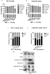
Figure 1
Figure 1
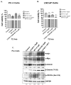
ATP cell viability assay: PC-3 (A) and LNCaP (B) cells were treated with MT-1 at varying concentrations (0.001–10 µM) for 72 h. MT-1 significantly decreased LNCaP cells growth in a dose dependent fashion at 1 and 10 µM concentrations of MT-1. PC-3 cells were resistant to low doses of MT-1 < 1 µM concentrations but the viability was significantly reduced at a higher dose of 10 µM MT-1 at 72 h (* indicates statistically significant compared to control, p < 0.05). Standard Error bars presented in the graph. The data suggest that MT-1 inhibits growth of LNCaP and PC-3 cells, albeit LNCaP cells being more sensitive to MT-1 inhibition at a lower concentration. Experiments were repeated in triplicate independently. MT-1 caused G0-G1 phase cell cycle arrest (C,D) in PC-3 and LNCaP cells, respectively, within 48 h in a dose dependent fashion. Western blot analysis of PC-3 cells with MT-1 at 0.1 and 0.5 µM concentrations for 24 h reduced and increased c-Myc and PrKD1 protein levels, respectively. Consistent with increased in PrKD expression, the known substrate phosphorylation of p-CDC25c (Ser-216), β-Catenin (T-120) and Snail (S-11) were increased from 1.11 to 2.5-fold, confirming an increase in PrKD kinase activity (E). The experiments were performed in triplicate independently. * p-value < 0.01.
Figure 2
Figure 2
ATP cell viability assay: PC-3 (A) and LNCaP (B) cells were treated with either MT-1, 3JC48-3 or 3JC48-3 and MT-1 at varying concentrations for 72 h. The combination of 3JC48-3 and MT-1 was most effective in reducing cell viability compared to control or any of the single drug treatments (* indicates statistically significant compared to control, p < 0.05). Standard Error bars presented in the graph. Western blot (C) Compared to control, treatment of PC-3 cells with the drugs for 24 h reduced and increased c-Myc and PrKD1 protein levels, respectively. Consistent with increased in PrKD1 expression, the known substrate phosphorylation of p-CDC25c (Ser-216), β-Catenin (T-120), and Snail (S-11) were increased confirming an increase in PrKD kinase activity. Like viability assay, the combination of 3JC48-3 and MT-1 was most effective compared to single-drug treatments. As PrKD1 expression has been shown to be repressed by androgens, we used MDV3100, an AR antagonist, as positive control to upregulate PrKD1 expression. The experiments were performed in triplicate independently. * p-value < 0.01, ** p-value < 0.001.
Figure 3
Figure 3
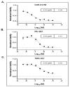
MT-1 compound effects on PC patient-derived 3D tumor spheroid culture: three-dimensional (3D) spheroid cell viability assay demonstrates MT-1 activity against three unique PC patient-derived tumor spheroids. (A) Metastatic castrate resistant PC GUR-017M patient tumor spheroid showing dose response growth inhibition at doses 0.5 µM to 10 µM, (B) metastatic castrate-resistant PC PRJ-88T patient tumor in spheroid showing dose response growth inhibition at doses 0.005 µM to 10 µM, and (C) metastatic castrate-resistant PC TMA-027 patient tumor in spheroid showing dose response growth inhibition at doses 0.01 µM to 10 µM of MT-1 compound. Standard Error bars presented in the graph.
Figure 4
Figure 4
c-Myc inhibition by MT-1 and/or 3JC48-3 inhibits tumor growth in c-Myc upregulated patient-derived PC xenograft mice model by 4 weeks (A). Western blot analysis of tumor samples treated with MT-1 increased expressions of PrKD1 along with increased substrate phosphorylation of Snail (S-11) and β-Catenin (T-120). GAPDH is loading control for protein (B).

References

    1. Asangani I.A., Dommeti V.L., Wang X., Malik R., Cieslik M., Yang R., Escara-Wilke J., Wilder-Romans K., Dhanireddy S., Engelke C. Therapeutic targeting of BET bromodomain proteins in castration-resistant prostate cancer. Nature. 2014;510:278–282. doi: 10.1038/nature13229. - DOI - PMC - PubMed
    1. Yu C., Niu X., Jin F., Liu Z., Jin C., Lai L. Structure-based inhibitor design for the intrinsically disordered protein c-Myc. Sci. Rep. 2016;6:22298. doi: 10.1038/srep22298. - DOI - PMC - PubMed
    1. Kumar D., Sharma N., Giri R. Therapeutic interventions of cancers using intrinsically disordered proteins as drug targets: C-Myc as model system. Cancer Inform. 2017;16:1176935117699408. doi: 10.1177/1176935117699408. - DOI - PMC - PubMed
    1. Kalkat M., Resetca D., Lourenco C., Chan P.-K., Wei Y., Shiah Y.-J., Vitkin N., Tong Y., Sunnerhagen M., Done S.J., et al. MYC Protein Interactome Profiling Reveals Functionally Distinct Regions that Cooperate to Drive Tumorigenesis. Mol. Cell. 2018;72:836–848.e7. doi: 10.1016/j.molcel.2018.09.031. - DOI - PubMed
    1. Whitfield A., Beaulieu M.-E., Soucek L. Strategies to Inhibit Myc and Their Clinical Applicability. Front. Cell Dev. Biol. 2017;5:10. doi: 10.3389/fcell.2017.00010. - DOI - PMC - PubMed