. 2023 Aug 5;15(15):3986.
doi: 10.3390/cancers15153986.

Plasma Exosome Proteins ILK1 and CD14 Correlated with Organ-Specific Metastasis in Advanced Gastric Cancer Patients

Affiliations

Plasma Exosome Proteins ILK1 and CD14 Correlated with Organ-Specific Metastasis in Advanced Gastric Cancer Patients

Chenfei Zhou et al. Cancers (Basel). .

Abstract

The exosome plays important roles in driving tumor metastasis, while the role of exosome proteins during organ-specific metastasis in gastric cancer has not been fully understood. To address this question, peripheral blood samples from 12 AGC patients with organ-specific metastasis, including distant lymphatic, hepatic and peritoneal metastasis, were collected to purify exosomes and to detect exosome proteins by Nano-HPLC-MS/MS. Gastric cancer cell lines were used for in vitro experiments. Peripheral blood sample and ascites sample from one patient were further analyzed by single-cell RNA sequencing. GO and KEGG enrichment analysis showed different expression proteins of hepatic metastasis were correlated with lipid metabolism. For peritoneal metastasis, actin cytoskeleton regulation and glycolysis/gluconeogenesis could be enriched. ILK1 and CD14 were correlated with hepatic and peritoneal metastasis, respectively. Overexpression of CD14 and ILK1 impacted the colony formation ability of gastric cancer and increased expression of Vimentin. CD14 derived from immune cells in malignant ascites correlated with high activation of chemokine- and cytokine-mediated signaling pathways. In summary, biological functions of plasma exosome proteins among AGC patients with different metastatic modes were distinct, in which ILK1 and CD14 were correlated with organ-specific metastasis.

Keywords: exosome; gastric cancer; organ-specific metastasis; tumor microenvironment.


Conflict of interest statement

The authors declare no conflict of interest.

Figures

Figure 1
Figure 1
DEPs of plasma exosomes detected in AGC patients with different organ-specific metastasis and GO analysis of DEPs. (A): Heatmap of DEPs’ average values in H group vs. L group and P group vs. L group. (B): Biological processes of GO analysis enriched by DEPs (***: FDR ≤ 0.001). (C): Molecular functions of GO analysis enriched by DEPs.
Figure 2
Figure 2
KEGG enrichment analysis of DEPs. (A): Signaling pathways of KEGG analysis enriched by DEPs. (B): Volcano plot of top-ranked DEPs from H group vs. L group and P group vs. L group.
Figure 3
Figure 3
Correlation of plasma ILK1 and CD14 with organ-specific metastasis. (A): Plasma level of ILK1 and CD14 in AGC patients with different metastatic modes. (B): ROC curve to identify cut-off values of ILK1 and CD14.
Figure 4
Figure 4
Overexpression of CD14 and ILK1 impacted biological behaviors in gastric cancer cells. (A): Expression of CD14 and ILK1 in MKN45 and NCI-N87 cells were detected by qRT-PCR and western blot. (B): Cell cycle analysis of CD14 and ILK1 overexpression cell models. (C): Representative images of colony formation assay. Colony size analysis was performed by ImageJ software. (D): Representative images of migration assay. (E): Expression of E-cadherin, Vimentin and Snail by western blot. Quantification data of western blot bands were analyzed by ImageJ software and compared with control groups. Original western blots have been presented in File S1.
Figure 5
Figure 5
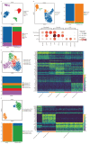
CD14 derived from subclusters of macrophages and monocytes in malignant ascites. (A): Cellular constitution of ascites sample and peripheral blood sample. (B): Cellular constitution of mononuclear phagocytes and cellular markers expression. (C): Subcluster analysis of macrophages in ascites samples. The heatmap illustrates the differential expression genes used to identify subcluster of macrophages. (D): Subcluster analysis of monocytes in ascites sample and peripheral blood sample. The heatmap illustrates the differential expression genes used to identify subcluster of monocytes.
Figure 6
Figure 6
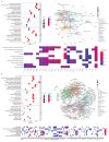
Biological functions analysis of high-expression DEGs of subclusters of macrophages and monocytes. (A): Dot plot and net plot of GO analysis of high-expression DEGs of Macrophage_FN1, and heat map of significantly enriched signaling pathways. (B): Dot plot and net plot of GO analysis of high-expression DEGs of ClassicalMono_CCL4L2, and heat map of significantly enriched signaling pathways.

References

    1. Shimizu D., Kanda M., Kodera Y. Emerging evidence of the molecular landscape specific for hematogenous metastasis from gastric cancer. World J. Gastrointest. Oncol. 2018;10:124–136. doi: 10.4251/wjgo.v10.i6.124. - DOI - PMC - PubMed
    1. Mikula-Pietrasik J., Uruski P., Tykarski A., Ksiazek K. The peritoneal “soil” for a cancerous “seed”: A comprehensive review of the pathogenesis of intraperitoneal cancer metastases. Cell. Mol. Life Sci. 2018;75:509–525. doi: 10.1007/s00018-017-2663-1. - DOI - PMC - PubMed
    1. Cunningham D., Starling N., Rao S., Iveson T., Nicolson M., Coxon F., Middleton G., Daniel F., Oates J., Norman A.R., et al. Capecitabine and oxaliplatin for advanced esophagogastric cancer. N. Engl. J. Med. 2008;358:36–46. doi: 10.1056/NEJMoa073149. - DOI - PubMed
    1. Psaila B., Lyden D. The metastatic niche: Adapting the foreign soil. Nat. Rev. Cancer. 2009;9:285–293. doi: 10.1038/nrc2621. - DOI - PMC - PubMed
    1. Jiang H., Yu D., Yang P., Guo R., Kong M., Gao Y., Yu X., Lu X., Fan X. Revealing the transcriptional heterogeneity of organ-specific metastasis in human gastric cancer using single-cell RNA Sequencing. Clin. Transl. Med. 2022;12:e730. doi: 10.1002/ctm2.730. - DOI - PMC - PubMed