

Features of Myocarditis: Morphological Differential Diagnosis in Post-COVID-19 Children
- PMID: 37568863
- PMCID: PMC10417761
- DOI: 10.3390/diagnostics13152499
Features of Myocarditis: Morphological Differential Diagnosis in Post-COVID-19 Children
Abstract
Myocarditis is characterized by dysfunction and destruction of cardiomyocytes, infiltrative inflammation, and development of fibrosis. Late diagnosis of myocarditis has been a serious global health problem, especially due to the spread of a new coronavirus infection. The aim of this review is to identify differences between myocarditis of viral etiology, including SARS-CoV-2 lesions, based on instrumental and pathomorphological findings.
Material and methods: We analyzed publications covering the period from December 2019 to May 2023, published in publicly accessible international databases ("Medline", "PubMed", "Scopus"), with queries for the keywords "myocarditis", "children", "cardiovascular inflammation", "COVID-19", "SARS-CoV-2", "severe acute respiratory syndrome coronavirus 2", "differential diagnosis".
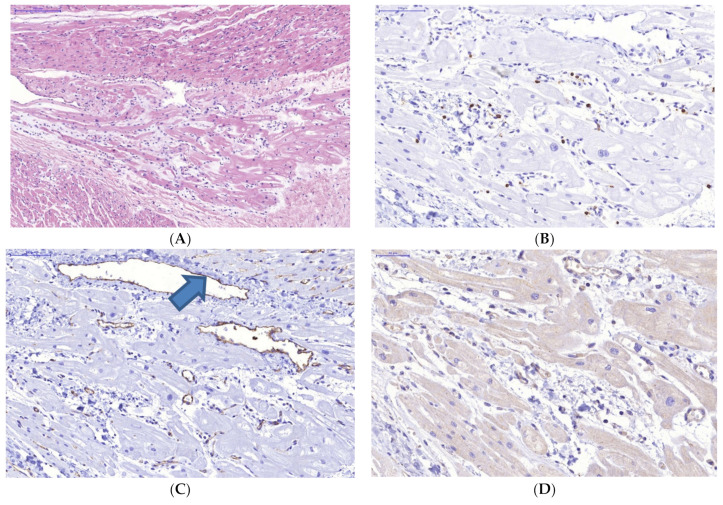
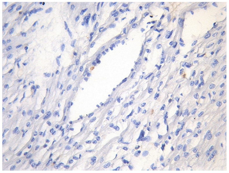
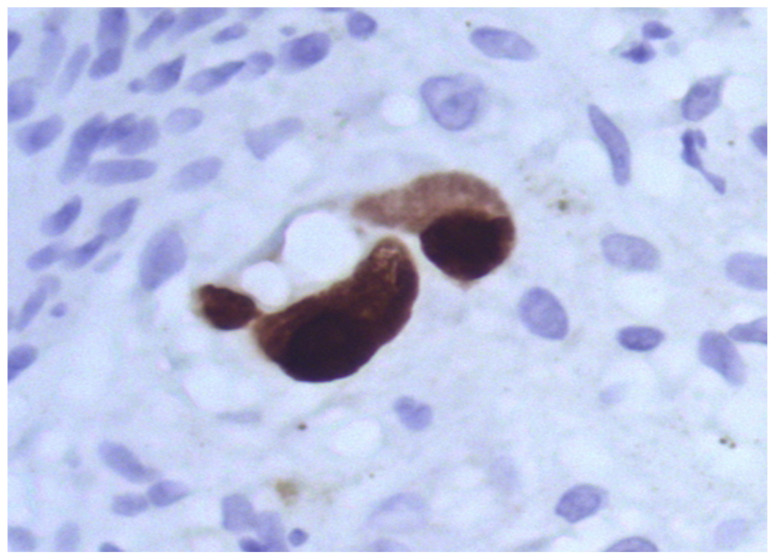
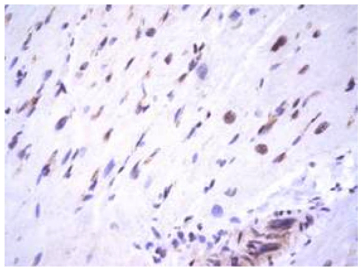
Results: It was found that no unambiguous morphological criteria for the diagnosis of myocarditis coupled to SARS-CoV-2 lesions were identified. However, the detected histopathological changes such as virus-associated degeneration, apoptosis, cardiomyocyte necrosis, moderate interstitial hyperemia, myocardial tissue oedema, and capillary endothelial cell dysfunction were the major markers of SARS-CoV-2 infection.
Conclusion: It is necessary further reconsider morphological criteria to diagnose SARS-CoV-2-caused myocarditis, rather than solely relying on detecting viral RNA by PCR as the sole evidence-based criterion. Similar issues accompany diagnostics of myocardial lesions associated with other viral infections. Evidence for an etiological diagnosis of myocarditis can be provided by a comprehensive analysis of the diagnostic criteria obtained, confirming virus exposure, followed by development of distinct clinical symptoms, MRI and CT changes, and morphological criteria.
Keywords: SARS-CoV-2 infection; heart transplantation; immunohistochemical and ultrastructural myocardial studies; virus transmission.
Conflict of interest statement
The authors declare no conflict of interest.
Figures
Similar articles
-
[Clinical and morphological characteristics of SARS-CoV-2-related myocarditis proven by the presence of viral RNA and proteins in myocardial tissue].Arkh Patol. 2021;83(4):5-13. doi: 10.17116/patol2021830415. Arkh Patol. 2021. PMID: 34278755 Russian.
-
Subacute and chronic post-covid myoendocarditis: clinical presentation, role of coronavirus persistence and autoimmune mechanisms.Kardiologiia. 2021 Jul 1;61(6):11-27. doi: 10.18087/cardio.2021.6.n1659. Kardiologiia. 2021. PMID: 34311684 English, Russian.
-
Clinically Suspected and Biopsy-Proven Myocarditis Temporally Associated with SARS-CoV-2 Infection.Annu Rev Med. 2022 Jan 27;73:149-166. doi: 10.1146/annurev-med-042220-023859. Epub 2021 Sep 10. Annu Rev Med. 2022. PMID: 34506211 Review.
-
Myocardial injury and COVID-19: Possible mechanisms.Life Sci. 2020 Jul 15;253:117723. doi: 10.1016/j.lfs.2020.117723. Epub 2020 Apr 28. Life Sci. 2020. PMID: 32360126 Free PMC article. Review.
-
Chronic biopsy proven post-COVID myoendocarditis with SARS-Cov-2 persistence and high level of antiheart antibodies.Clin Cardiol. 2022 Sep;45(9):952-959. doi: 10.1002/clc.23886. Epub 2022 Jul 19. Clin Cardiol. 2022. PMID: 35855554 Free PMC article.
Cited by
-
Clinical Presentation and Outcomes of Myocarditis Among the COVID-19 Pediatric Population: A Review of 100 Cases.Cureus. 2024 Sep 21;16(9):e69885. doi: 10.7759/cureus.69885. eCollection 2024 Sep. Cureus. 2024. PMID: 39439599 Free PMC article.
-
The Promising Role of Speckle-tracking Echocardiography in Detecting the Patchy Nature of Myocarditis, Insights from Case Series in the Largest Pediatrics Tertiary Center of UAE.Heart Views. 2024 Jul-Sep;25(3):157-161. doi: 10.4103/heartviews.heartviews_54_24. Epub 2025 Jan 4. Heart Views. 2024. PMID: 40028244 Free PMC article.
-
Are Endomyocardial Ventricular Biopsies Useful for Assessing Myocardial Fibrosis?J Clin Med. 2024 May 31;13(11):3275. doi: 10.3390/jcm13113275. J Clin Med. 2024. PMID: 38892986 Free PMC article.
References
-
- WHO . Coronavirus Disease (COVID-19) Pandemic. WHO; Geneva, Switzerland: 2020. [(accessed on 8 June 2023)]. Available online: https://www.who.int/emergencies/diseases/novel-coronavirus-2019.
-
- WHO Coronavirus (COVID-19) Dashboard. [(accessed on 10 December 2022)]. Available online: https://covid19.who.int/?mapFilter=deaths.
-
- WHO Coronavirus Disease (COVID-19): Schools. [(accessed on 10 December 2022)]. Available online: https://www.who.int/news-room/questions-and-answers/item/coronavirus-dis....
Publication types
Grants and funding
LinkOut - more resources
Full Text Sources
Miscellaneous
