

Retinal Photoreceptors and Microvascular Changes in the Assessment of Diabetic Retinopathy Progression: A Two-Year Follow-Up Study
- PMID: 37568876
- PMCID: PMC10417253
- DOI: 10.3390/diagnostics13152513
Retinal Photoreceptors and Microvascular Changes in the Assessment of Diabetic Retinopathy Progression: A Two-Year Follow-Up Study
Abstract
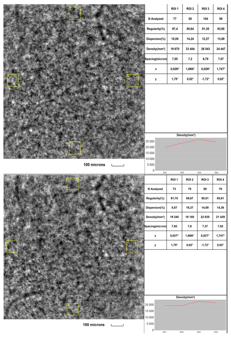
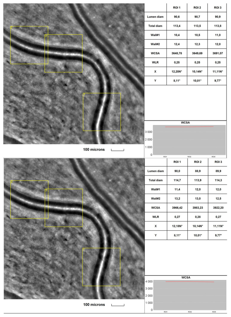
Background: With the increasing global incidence of diabetes mellitus (DM), diabetic retinopathy (DR) has become one of the leading causes of blindness in developed countries. DR leads to changes in retinal neurons and microcirculation. Rtx1TM (Imagine Eyes, Orsay, France) is a retinal camera that allows histological visualisations of cones and retinal microcirculation throughout the DM duration.
Objective: This study aimed to analyse the cones and retinal microvascular changes in 50 diabetic individuals and 18 healthy volunteers. The patients participated in the initial visit and two follow-up appointments, one and two years after the study, beginning with Rtx1TM image acquisition, visual acuity assessment, macular OCT scans and blood measurements.
Results: The study revealed significant differences in the cone density, mosaic arrangement and vascular morphology between healthy and diabetic patients. The final measurements showed decreased photoreceptor and microvascular parameters in the DR group compared with the control group. Furthermore, in the 2-year follow-up, both groups' Rtx1TM-acquired morphological changes were statistically significant.
Conclusions: Rtx1TM technology was successfully used as a non-invasive method of photoreceptors and retinal vasculature assessment over time in patients with diabetic retinopathy. The study revealed a trend toward more vascular morphological changes occurring over time in diabetic patients.
Keywords: adaptive optics; cone morphology; diabetic retinopathy; retinal microcirculation; rtx-1 technology.
Conflict of interest statement
The authors declared no conflict of interest. None of the authors had any financial interest concerning the presented subject.
Figures
References
-
- Saeedi P., Petersohn I., Salpea P., Malanda B., Karuranga S., Unwin N., Colagiuri S., Guariguata L., Motala A.A., Ogurtsova K., et al. Global and regional diabetes prevalence estimates for 2019 and projections for 2030 and 2045: Results from the International Diabetes Federation Diabetes Atlas, 9th edition. Diabetes Res. Clin. Pract. 2019;157:107843. doi: 10.1016/j.diabres.2019.107843. - DOI - PubMed
-
- Wilkinson C.P., Ferris F.L., Klein R.E., Lee P.P., Agardh C.D., Davis M., Dils D., Kmapik A., Pararajsegaram R., Verdaguer J.T., et al. Proposed international clinical diabetic retinopathy and diabetic macular oedema disease severity scales. Ophthalmology. 2003;110:1677–1682. doi: 10.1016/S0161-6420(03)00475-5. - DOI - PubMed
LinkOut - more resources
Full Text Sources
