

Echocardiographic Evaluation of Aortic Stenosis: A Comprehensive Review
- PMID: 37568890
- PMCID: PMC10417789
- DOI: 10.3390/diagnostics13152527
Echocardiographic Evaluation of Aortic Stenosis: A Comprehensive Review
Abstract
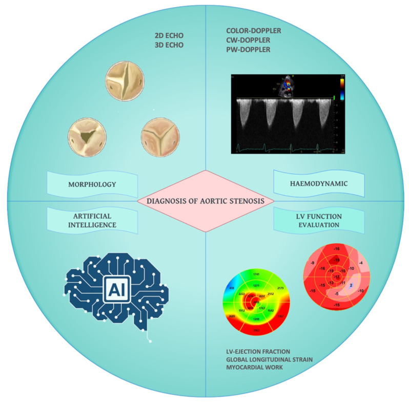
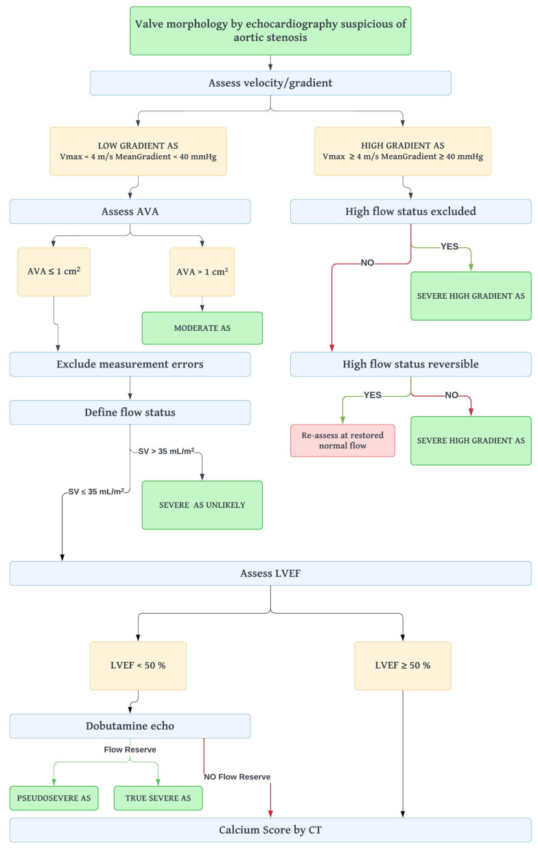
Echocardiography represents the most important diagnostic tool in the evaluation of aortic stenosis. The echocardiographic assessment of its severity should always be performed through a standardized and stepwise approach in order to achieve a comprehensive evaluation. The latest technical innovations in the field of echocardiography have improved diagnostic accuracy, guaranteeing a better and more detailed evaluation of aortic valve anatomy. An early diagnosis is of utmost importance since it shortens treatment delays and improves patient outcomes. Echocardiography plays a key role also in the evaluation of all the structural changes related to aortic stenosis. Detailed evaluation of subtle and subclinical changes in left ventricle function has a prognostic significance: scientific efforts have been addressed to identify the most accurate global longitudinal strain cut-off value able to predict adverse outcomes. Moreover, in recent years the role of artificial intelligence is increasingly emerging as a promising tool able to assist cardiologists in aortic stenosis screening and diagnosis, especially by reducing the rate of aortic stenosis misdiagnosis.
Keywords: aortic stenosis; aortic stenosis grading; artificial intelligence; cardiac damage; echocardiography; multiparametric approach.
Conflict of interest statement
The authors declare no conflict of interest.
Figures
References
-
- Durko A.P., Osnabrugge R.L., van Mieghem N.M., Milojevic M., Mylotte D., Nkomo V.T., Pieter Kappetein A. Annual Number of Candidates for Transcatheter Aortic Valve Implantation per Country: Current Estimates and Future Projections. Eur. Heart J. 2018;39:2635–2642. doi: 10.1093/eurheartj/ehy107. - DOI - PubMed
-
- Nicoara A., Skubas N., Ad N., Finley A., Hahn R.T., Mahmood F., Mankad S., Nyman C.B., Pagani F., Porter T.R., et al. Guidelines for the Use of Transesophageal Echocardiography to Assist with Surgical Decision-Making in the Operating Room: A Surgery-Based Approach: From the American Society of Echocardiography in Collaboration with the Society of Cardiovascular Anesthesiologists and the Society of Thoracic Surgeons. J. Am. Soc. Echocardiogr. 2020;33:692–734. - PubMed
-
- Baumgartner H., Hung J., Bermejo J., Chambers J.B., Edvardsen T., Goldstein S., Lancellotti P., LeFevre M., Miller F., Otto C.M. Recommendations on the Echocardiographic Assessment of Aortic Valve Stenosis: A Focused Update from the European Association of Cardiovascular Imaging and the American Society of Echocardiography. J. Am. Soc. Echocardiogr. 2017;30:372–392. doi: 10.1016/j.echo.2017.02.009. - DOI - PubMed
Publication types
LinkOut - more resources
Full Text Sources
