Review
. 2023 Jul 25;24(15):11913.
doi: 10.3390/ijms241511913.

Microtubule-Associated Serine/Threonine (MAST) Kinases in Development and Disease

Affiliations
Review

Microtubule-Associated Serine/Threonine (MAST) Kinases in Development and Disease

Marie Rumpf et al. Int J Mol Sci. .

Abstract

Microtubule-Associated Serine/Threonine (MAST) kinases represent an evolutionary conserved branch of the AGC protein kinase superfamily in the kinome. Since the discovery of the founding member, MAST2, in 1993, three additional family members have been identified in mammals and found to be broadly expressed across various tissues, including the brain, heart, lung, liver, intestine and kidney. The study of MAST kinases is highly relevant for unraveling the molecular basis of a wide range of different human diseases, including breast and liver cancer, myeloma, inflammatory bowel disease, cystic fibrosis and various neuronal disorders. Despite several reports on potential substrates and binding partners of MAST kinases, the molecular mechanisms that would explain their involvement in human diseases remain rather obscure. This review will summarize data on the structure, biochemistry and cell and molecular biology of MAST kinases in the context of biomedical research as well as organismal model systems in order to provide a current profile of this field.

Keywords: MAST kinase; cell signaling; protein phosphorylation.


Conflict of interest statement

The authors declare no conflict of interest.

Figures

Figure 1
Figure 1
Domain arrangement of human MAST kinases. This cartoon demonstrates the domain composition and arrangement of the four family members of MAST kinases in humans. From N- to C-terminus, the DUF 1908 domain is followed by the serine/threonine kinase domain (drawn without its C-terminal extension) and the PDZ domain in the C-terminal half of the protein.
Figure 2
Figure 2
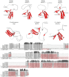
Primary sequence comparison and structural comparison of DUF1908 domains in selected MAST kinases. Top panel: AlphaFold predicted 3D models of DUF1908 of MAST kinases in humans, Drosophila (D.) melanogaster, Caenorhabditis (C.) elegans and Hydra (H.) vulgaris [13,14,15]. Alpha helices are shown in red. The 3D structures were oriented and labeled using the PyMol software (Schrödinger, L.; DeLano, W. PyMOL [Internet]. 2020. Available online: http://www.pymol.org/pymol; accessed on 5 December 2022). Lower panel: Multiple amino acid sequence alignment of DUF1908 of MAST kinases in different species using MAFFT (Snapgen; GSL Biotec, San Diego, CA, USA). The helices are marked in red. Conserved amino acids are highlighted in gray and dark red, respectively. A threshold of 55% was chosen for consensus. The bars above the consensus sequence indicate the degree of conservation; the darker and higher the bar, the higher the conservation. The sequences and domain annotations of the proteins were obtained from InterPro [16].
Figure 3
Figure 3
Primary sequence comparison and structural comparison of PDZ domains in selected MAST kinases. AlphaFold-predicted 3D models of PDZ domains of MAST kinases in humans, Drosophila (D.) melanogaster, Caenorhabditis (C.) elegans, and Hydra (H.) vulgaris [13,14,15]. Alpha helices are shown in red, and beta sheets in blue color. The 3D structures were oriented and labeled using the PyMol software (Schrödinger, LLC, New York, NY, USA). The structure of all four human MAST kinases was also solved experimentally (MAST1: https://doi.org/10.2210/pdb3ps4/pdb; MAST2: https://doi.org/10.2210/pdb2kyl/pdb; MAST3: https://doi.org/10.2210/pdb3khf/pdb; MAST4: https://doi.org/10.2210/pdb2w7r/pdb) (accessed on 14 July 2023). The structural models based on AlphaFold and based on experimental data do not exhibit appreciable differences. Lower panel: Multiple protein amino acid sequence alignment of PDZ domains of MAST kinases in different species using MAFFT (Snapgen; GSL Biotec). For further detail see the legend of Figure 2.
Figure 4
Figure 4
Structure models and sequence comparison of the MAST kinase domains in different species. Top panels: AlphaFold-prediction models of the kinase domain structure in humans, Drosophila (D.) melanogaster, Caenorhabditis (C.) elegans, and Hydra (H.) vulgaris [13,14,15]. The alpha helices are shown in red, and beta sheets in blue. The 3D structures were oriented and labeled using PyMol (Schrödinger, LLC). Lower panel: Multiple protein amino acid sequence alignment of MAST kinase domains of different species. Alignment was performed using MAFFT of the Snapgen software (GSL Biotec). For further detail see the legend of Figure 2.
Figure 5
Figure 5
Sequence comparison of the AGC-kinase C-terminal region in MAST kinases of different species. Multiple protein amino acid sequence alignments of AGC-kinase C-terminal regions of MAST kinases in humans, Drosophila (D.) melanogaster and Hydra (H.) vulgaris. Alignment was performed using MAFFT (Snapgene; GSL Biotec). Predicted alpha-helical domains are marked in red and conserved amino acids are marked in grey or dark red.
Figure 6
Figure 6
Phylogenetic analyses of MAST kinases. Maximum likelihood phylogenetic tree showing the relation of MAST, MAST-like and putative MAST kinases with similar protein domain architecture from simple organisms like Trichoplax adhaerens, Hydra vulgaris to higher organisms such as Homo sapiens and Mus musculus. The protein domain annotations were identified by SMART [113] and the alignment was performed by using MAFFT version 7 [114] and RAxML was used for phylogenetic analysis [115]. All bootstrap values are shown, and the tree image was drawn using the iTOL web server [111,112].

References

    1. Manning G., Whyte D.B., Martinez R., Hunter T., Sudarsanam S. The protein kinase complement of the human genome. Science. 2002;298:1912–1934. doi: 10.1126/science.1075762. - DOI - PubMed
    1. Richardson C.J., Gao Q., Mitsopoulous C., Zvelebil M., Pearl L.H., Pearl F.M. MoKCa database--mutations of kinases in cancer. Nucleic Acids Res. 2009;37:D824–D831. doi: 10.1093/nar/gkn832. - DOI - PMC - PubMed
    1. Hanks S.K., Quinn A.M., Hunter T. The protein kinase family: Conserved features and deduced phylogeny of the catalytic domains. Science. 1988;241:42–52. doi: 10.1126/science.3291115. - DOI - PubMed
    1. Hanks S.K., Hunter T. Protein kinases 6. The eukaryotic protein kinase superfamily: Kinase (catalytic) domain structure and classification. FASEB J. 1995;9:576–596. doi: 10.1096/fasebj.9.8.7768349. - DOI - PubMed
    1. Pearce L.R., Komander D., Alessi D.R. The nuts and bolts of AGC protein kinases. Nat. Rev. Mol. Cell Biol. 2010;11:9–22. doi: 10.1038/nrm2822. - DOI - PubMed

LinkOut - more resources