

T-Cell Prolymphocytic Leukemia: Diagnosis, Pathogenesis, and Treatment
- PMID: 37569479
- PMCID: PMC10419310
- DOI: 10.3390/ijms241512106
T-Cell Prolymphocytic Leukemia: Diagnosis, Pathogenesis, and Treatment
Abstract
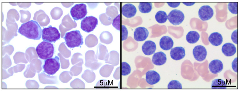
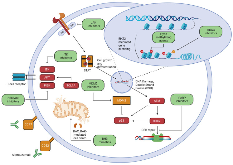
T-cell prolymphocytic leukemia (T-PLL) is a rare and aggressive neoplasm of mature T-cells. Most patients with T-PLL present with lymphocytosis, anemia, thrombocytopenia, and hepatosplenomegaly. Correct identification of T-PLL is essential because treatment for this disease is distinct from that of other T-cell neoplasms. In 2019, the T-PLL International Study Group (TPLL-ISG) established criteria for the diagnosis, staging, and assessment of response to treatment of T-PLL with the goal of harmonizing research efforts and supporting clinical decision-making. T-PLL pathogenesis is commonly driven by T-cell leukemia 1 (TCL1) overexpression and ATM loss, genetic alterations that are incorporated into the TPLL-ISG diagnostic criteria. The cooperativity between TCL1 family members and ATM is seemingly unique to T-PLL across the spectrum of T-cell neoplasms. The role of the T-cell receptor, its downstream kinases, and JAK/STAT signaling are also emerging themes in disease pathogenesis and have obvious therapeutic implications. Despite improved understanding of disease pathogenesis, alemtuzumab remains the frontline therapy in the treatment of naïve patients with indications for treatment given its high response rate. Unfortunately, the responses achieved are rarely durable, and the majority of patients are not candidates for consolidation with hematopoietic stem cell transplantation. Improved understanding of T-PLL pathogenesis has unveiled novel therapeutic vulnerabilities that may change the natural history of this lymphoproliferative neoplasm and will be the focus of this concise review.
Keywords: ATM; BH3 mimetic; JAK; STAT; T-cell receptor; T-prolymphocytic leukemia (T-PLL); TCL1A; alemtuzumab.
Conflict of interest statement
The authors declare that the research was conducted in the absence of any commercial or financial relationships that could be construed as a potential conflict of interest.
Figures
References
-
- Staber P.B., Herling M., Bellido M., Jacobsen E.D., Davids M.S., Kadia T.M., Shustov A., Tournilhac O., Bachy E., Zaja F., et al. Consensus Criteria for Diagnosis, Staging, and Treatment Response Assessment of T-Cell Prolymphocytic Leukemia. Blood. 2019;134:1132–1143. doi: 10.1182/blood.2019000402. - DOI - PMC - PubMed
-
- Alaggio R., Amador C., Anagnostopoulos I., Attygalle A.D., de Oliveira Araujo I.B., Berti E., Bhagat G., Borges A.M., Boyer D., Calaminici M., et al. The 5th Edition of the World Health Organization Classification of Haematolymphoid Tumours: Lymphoid Neoplasms. Leukemia. 2022;36:1720–1748. doi: 10.1038/s41375-022-01620-2. - DOI - PMC - PubMed
Publication types
MeSH terms
Substances
Grants and funding
LinkOut - more resources
Full Text Sources
Research Materials
Miscellaneous
