. 2023 Jul 31;24(15):12270.
doi: 10.3390/ijms241512270.

Brainstem Modulates Parkinsonism-Induced Orofacial Sensorimotor Dysfunctions

Affiliations

Brainstem Modulates Parkinsonism-Induced Orofacial Sensorimotor Dysfunctions

Glauce Crivelaro Nascimento et al. Int J Mol Sci. .

Abstract

Parkinson's Disease (PD), treated with the dopamine precursor l-3,4-dihydroxyphenylalanine (L-DOPA), displays motor and non-motor orofacial manifestations. We investigated the pathophysiologic mechanisms of the lateral pterygoid muscles (LPMs) and the trigeminal system related to PD-induced orofacial manifestations. A PD rat model was produced by unilateral injection of 6-hydroxydopamine into the medial forebrain bundle. Abnormal involuntary movements (dyskinesia) and nociceptive responses were determined. We analyzed the immunodetection of Fos-B and microglia/astrocytes in trigeminal and facial nuclei and morphological markers in the LPMs. Hyperalgesia response was increased in hemiparkinsonian and dyskinetic rats. Hemiparkinsonism increased slow skeletal myosin fibers in the LPMs, while in the dyskinetic ones, these fibers decreased in the contralateral side of the lesion. Bilateral increased glycolytic metabolism and an inflammatory muscle profile were detected in dyskinetic rats. There was increased Fos-B expression in the spinal nucleus of lesioned rats and in the motor and facial nucleus in L-DOPA-induced dyskinetic rats in the contralateral side of the lesion. Glial cells were increased in the facial nucleus on the contralateral side of the lesion. Overall, spinal trigeminal nucleus activation may be associated with orofacial sensorial impairment in Parkinsonian rats, while a fatigue profile on LPMs is suggested in L-DOPA-induced dyskinesia when the motor and facial nucleus are activated.

Keywords: brain function; muscle biology; neuroscience/neurobiology; neurotoxicity; pain.


Conflict of interest statement

The authors declare no conflict of interest. The funders had no role in the design of the study; in the collection, analyses, or interpretation of data; in the writing of the manuscript; or in the decision to publish the results.

Figures
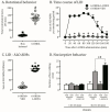
Figure 1
Figure 1
Behavioral and nociceptive alterations induced by 6-OHDA and L-DOPA chronic treatment. (A): The microinjection of 6-OHDA in the unilateral nigrostriatal pathway induced robust contralateral rotational behavior after apomorphine challenge; # p < 0.001, comparing 6-OHDA lesioned rats to no-lesioned ones. (B): Time course of development of abnormal involuntary movements (AIMs) by chronic administration of L-DOPA to 6-OHDA-lesioned; * p < 0.001, comparing lesioned rats treated with L-DOPA or its vehicle. (C): Sum of AIMs during 180 min; * p < 0.001, comparing lesioned rats treated with L-DOPA or its vehicle. (D): Nociceptive behavior detected by formalin test. The duration of face rubbing is quantified in two phases of the analysis (I and II); # p < 0.001, comparing lesioned rats (6-OHDA) with non-lesioned ones (VEH); * p < 0.001, comparing lesioned rats treated with L-DOPA (dyskinetic) or its vehicle. Data are mean ± SEM. Two-way ANOVA revealed a main effect of experimental condition (F(2,19) = 11.7), treatment (F(3,19) = 9.78) and an interaction (F(3,19) = 19.2; p < 0.05).
Figure 2
Figure 2
Injury in the lateral pterygoid muscle due to L-DOPA-induced dyskinesia. Lateral pterygoid muscles (LPM) from lesioned and non-lesioned rats treated with L-DOPA or its vehicle were processed immunohistochemically for Miosin (A,B) and histological SDH enzyme and inflammatory infiltrate (C,D,G) analysis. (A,B): Representative photomicrographs from LPM identifying Myosin fibers in non-lesioned (VEH/L-DOPA) and lesioned rats (6-OHDA/L-DOPA) treated with L-DOPA (20×). (C,D): Representative photomicrographs identifying glycolytic fibers through SDH histochemical reaction in non-lesioned (VEH/L-DOPA) and lesioned rats (6-OHDA/L-DOPA) treated with L-DOPA (40×). (E): Quantification of Myosin fibers on ipsilateral (right) and contralateral (left) LPM; # p < 0.001, comparing 6-OHDA lesioned rats to non-lesioned ones; * p < 0.001, comparing lesioned rats treated with L-DOPA (dyskinetic) or its vehicle. The two-way ANOVA revealed a main effect of treatment (ipsilateral; F(1,19) = 14.47; p < 0.05 contralateral; F(1,19) = 10.78; p = 0.001). (F): Quantification of glycolytic fibers on ipsilateral (right) and contralateral (left) LPM; # p < 0.05, comparing lesioned rats treated with L-DOPA (dyskinetic) with all other experimental groups. Data are mean ± SEM. The two-way ANOVA revealed a main effect of treatment (contralateral; F(1,19) = 11.88). (G): Representative photomicrographs from LPM staining with hematoxylin and eosin in four groups used in this study -lesioned (6-OHDA+VEH) and non-lesioned (VEH+VEH) treated with L-DOPA (6-OHDA+L-DOPA) or its vehicle (6-OHDA+VEH). Bar represents 100 μm. Non-lesioned or lesioned groups treated with vehicle presented muscular fibers with polygonal aspect and varied size, with peripheral nuclei. Within each fascicle, the endomysium has uniform spacing with the presence of capillaries and cells in the conjunctiva. Isolated points of endomysial thickening were evidenced near the surface of an irregularly shaped muscle fiber. In the experimental group with LID, however, was observed the endomysium with inflammatory cells (macrophage and neutrophil).
Figure 3
Figure 3
Activation of the Trigeminal system by L-DOPA in 6-OHDA-lesioned rats with dyskinesia. Two trigeminal nucleus (Motor: −8.80 to −9.80 and Spinal: −10.30 to −11.60) and the Facial nucleus (−10.30 to −11.60) were analyzed by Fos-B immunohistochemistry to the evaluation of their neuronal activation in non-lesioned (VEH/L-DOPA), and lesioned rats (6-OHDA/L-DOPA) treated with L-DOPA. Schematic representation of (A) motor nucleus (Mo5 bregma: −8.80), (C) facial nucleus (7N, bregma: −10.80) and (E) spinal nucleus (Sp5O, bregma: −10.80) from Paxinos and Watson (2006). Photomicrographs showing Fos-B-is in Mo5 (A’,A”); 7N (C’,C”) and Sp5O (E’,E”) (20×). Graphics showing the number of Fos-B-in cells induced by L-DOPA treatment in lesioned rats Mo5 (B); 7N (D) and Sp5O (F) * p < 0.05, compared with all other experimental groups. The two-way ANOVA revealed a main effect of treatment (ipsilateral; F(1,19) = 13.66).
Figure 4
Figure 4
Neuroplasticity in the Facial nucleus through glial hyperactivity induced by L-DOPA-induced dyskinesia. Astrocyte (GFAP) and Microglia (OX-42) immunohistochemical in Motor (Mo5), Spinal (Sp5O) and Facial (7N) nucleus. (AD): Representative photomicrographs of GFAP and OX-42 staining in 7N (Bregma −10.80) in non-lesioned (VEH/L-DOPA) and lesioned rats (6-OHDA/L-DOPA) treated with L-DOPA (20×). (E): Quantification of GFAP (optic density) in 7N; * p < 0.05, comparing lesioned rats treated with L-DOPA (dyskinetic) with all other experimental groups. (F): Quantification of OX-42 (optic density) in 7N; * p < 0.05, comparing lesioned rats treated with L-DOPA (dyskinetic) with all other experimental groups. (G): Quantification of GFAP and OX-42 (optic density) in Mo5 and Sp5O; there was no statistical difference between the groups. Data are mean ± SEM. Bar represents 100 um. The two-way ANOVA revealed a main effect of treatment (ipsilateral; F(1,19) = 9.64; p = 0.04).
Figure 5
Figure 5
Experimental Design: Three weeks after the 6-OHDA lesion surgery, 48 animals were exposed to the apomorphine rotational test. Twenty-four of them constituted lesioned rats, and the other 24 were characterized as non-lesioned ones. Subsequently, 12 rats from each of the two groups were randomly assigned to be exposed to the drug treatment and the 12 remaining were chronically submitted to L-DOPA treatment. L-DOPA treatment started two days after the apomorphine rotational test. After chronic treatment with L-DOPA, rats were divided into experimental groups based on their dyskinesia score (AIMs scores). Moreover, on this last day, the experimental groups were submitted to a nociceptive behavior test, the animals were sacrificed, and lateral pterygoid muscle and brainstem were dissected for analyses (Miosin, SDH, HE, Fos-B and Glia). The nociceptive test and euthanasia were performed three hours after the last L-DOPA or vehicle administration. This experimental procedure was performed for immunohistochemistry analysis (n = 48 perfused animals) and muscle techniques (n = 48 non-perfused animals).

References

    1. Abdelhamid R.E., Sluka K.A. ASICs Mediate Pain and Inflammation in Musculoskeletal Diseases. Physiology. 2015;30:449–459. doi: 10.1152/physiol.00030.2015. - DOI - PMC - PubMed
    1. Uchida T., Adachi K., Fujita S., Lee J., Gionhaku N., Cools A.R., Koshikawa N. Role of GABA(A) receptors in the retrorubral field and ventral pallidum in rat jaw movementselicited by dopaminergic stimulation of the nucleus accumbens shell. Eur. J. Pharmacol. 2005;510:39–47. doi: 10.1016/j.ejphar.2005.01.012. - DOI - PubMed
    1. van Stiphout M.A.E., Marinus J., van Hilten J.J., Lobbezoo F., de Baat C. Oral Health of Parkinson’s Disease Patients: A Case-Control Study. Park. Dis. 2018;2018:9315285. doi: 10.1155/2018/9315285. - DOI - PMC - PubMed
    1. Fahn S. How do you treat motor complications in Parkinson’s disease: Medicine, surgery, or both? Ann. Neurol. 2008;64:56–64. doi: 10.1002/ana.21453. - DOI - PubMed
    1. Koshikawa N., Fujita S., Adachi K. Behavioral pharmacology of orofacial movement disorders. Int. Rev. Neurobiol. 2011;97:1–38. - PubMed