. 2023 Jul 31;24(15):12282.
doi: 10.3390/ijms241512282.

Polynucleotides Suppress Inflammation and Stimulate Matrix Synthesis in an In Vitro Cell-Based Osteoarthritis Model

Affiliations

Polynucleotides Suppress Inflammation and Stimulate Matrix Synthesis in an In Vitro Cell-Based Osteoarthritis Model

Sree Samanvitha Kuppa et al. Int J Mol Sci. .

Abstract

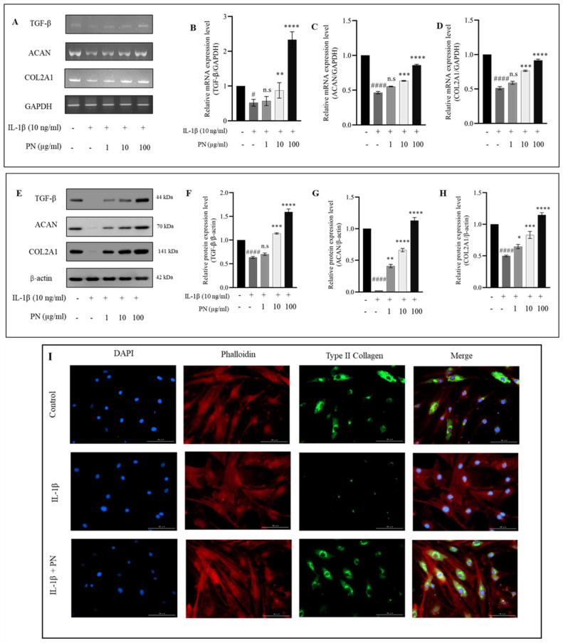
Osteoarthritis (OA) is characterized by degeneration of the joint cartilage, inflammation, and a change in the chondrocyte phenotype. Inflammation also promotes cell hypertrophy in human articular chondrocytes (HC-a) by activating the NF-κB pathway. Chondrocyte hypertrophy and inflammation promote extracellular matrix degradation (ECM). Chondrocytes depend on Smad signaling to control and regulate cell hypertrophy as well as to maintain the ECM. The involvement of these two pathways is crucial for preserving the homeostasis of articular cartilage. In recent years, Polynucleotides Highly Purified Technology (PN-HPT) has emerged as a promising area of research for the treatment of OA. PN-HPT involves the use of polynucleotide-based agents with controlled natural origins and high purification levels. In this study, we focused on evaluating the efficacy of a specific polynucleotide sodium agent, known as CONJURAN, which is derived from fish sperm. Polynucleotides (PN), which are physiologically present in the matrix and function as water-soluble nucleic acids with a gel-like property, have been used to treat patients with OA. However, the specific mechanisms underlying the effect remain unclear. Therefore, we investigated the effect of PN in an OA cell model in which HC-a cells were stimulated with interleukin-1β (IL-1β) with or without PN treatment. The CCK-8 assay was used to assess the cytotoxic effects of PN. Furthermore, the enzyme-linked immunosorbent assay was utilized to detect MMP13 levels, and the nitric oxide assay was utilized to determine the effect of PN on inflammation. The anti-inflammatory effects of PN and related mechanisms were investigated using quantitative PCR, Western blot analysis, and immunofluorescence to examine and analyze relative markers. PN inhibited IL-1β induced destruction of genes and proteins by downregulating the expression of MMP3, MMP13, iNOS, and COX-2 while increasing the expression of aggrecan (ACAN) and collagen II (COL2A1). This study demonstrates, for the first time, that PN exerted anti-inflammatory effects by partially inhibiting the NF-κB pathway and increasing the Smad2/3 pathway. Based on our findings, PN can potentially serve as a treatment for OA.

Keywords: chondrocytes; extracellular matrix; hypertrophy; inflammation; osteoarthritis; polynucleotide.


Conflict of interest statement

The authors declare no conflict of interest.

Figures

Figure 1
Figure 1
HC-a cell viability was examined in response to various concentrations of IL−1β after stimulation for (A) 24 and (B) 48 h. Additionally, (C) IL−1β concentration-dependent mRNA expressions of MMP13 and COL2A1 were evaluated using RT-PCR, and (D,E) quantitative analysis was performed. Following identification of the optimal IL−1β concentration (10 ng/mL), (F) the expression levels and (G,H) quantitative analyses of MMP13 and COL2A1 were evaluated at different time points (0, 3, 6, 9, 12, 24, and 48 h) using RT-PCR. The data, presented as the mean ± standard deviation (n = 3), were analyzed for statistical significance using the following significance notations: n.s (no significance), * p < 0.05, ** p < 0.01, *** p < 0.001, and **** p < 0.0001 compared to the control group (unstimulated cells).
Figure 2
Figure 2
(A) Cell viability of HC-a chondrocytes in response to varying concentrations of polynucleotide (PN). The viability of HC-a chondrocytes was measured after exposure to different concentrations of PN. (B) Cell viability of IL−1β-treated HC-a chondrocytes in response to PN. The cell viability of HC-a chondrocytes, pre-treated with IL−1β, was assessed following exposure to PN. Data are presented as mean ± standard deviation (n = 3). Statistical analysis revealed significant differences, with the following symbols denoting significance values: n.s (no significance); #### p < 0.0001 compared to the control group (unstimulated cells); * p < 0.05, ** p < 0.01, *** p < 0.001, and **** p < 0.0001 compared to the IL−1β-treated group.
Figure 3
Figure 3
(A) Effect of polynucleotide (PN) treatment on the expression of MMP13 in IL−1β-induced HC-a. MMP13 levels were assessed using an enzyme-linked immunosorbent assay (ELISA) in HC-a treated with IL−1β and PN. (B) Effect of PN treatment on IL−1β-induced production of nitric oxide (NO) in HC-a chondrocytes. Nitric oxide levels were measured using a nitric oxide assay in HC-a chondrocytes treated with IL−1β and PN. Data are presented as the mean ± standard deviation (n = 3). Statistical analysis revealed significant differences, with the following symbols denoting significance values: #### p < 0.0001 compared to the control group (unstimulated cells); n.s (no significance); * p < 0.05, ** p < 0.01, and **** p < 0.0001 compared to the IL−1β-treated group.
Figure 4
Figure 4
The effects of polynucleotide (PN) on inflammation and hypertrophy in IL−1β-induced HC-a. (A) Gene expression levels of MMP3, MMP13, iNOS, and COX2 were analyzed using RT-PCR. (BE) Quantitative analysis of MMP3, MMP13, iNOS, and COX2 gene expression. (F) Protein expression levels of MMP3, MMP13, iNOS, and COX2 were determined through Western blot analysis. (G–J) Quantitative analysis of MMP3, MMP13, iNOS, and COX2 protein expression. (K) Immunofluorescence staining was performed to visualize the expression and localization of iNOS (bars = 100 μm; original magnification × 20). Data are presented as the mean ± standard deviation (n = 3). Statistical analysis revealed significant differences, with the following symbols denoting significance values: n.s (no significance); ### p < 0.001, #### p < 0.0001 compared to the control group (unstimulated cells); and * p < 0.05, ** p < 0.01, *** p < 0.001, and **** p < 0.0001 compared to the IL−1β-treated group.
Figure 5
Figure 5
PN reduces IL−1β-induced HC-a inflammation through the NF-κB signaling pathway. (A) Whole cell lysate protein expression and (BE) quantitative analyses of total and phosphorylated forms of p65 and IκBα protein expression levels. Effect of PN on the expression of pp65 protein in HC-a cells stimulated by IL−1β in the presence of the 5HPP-33 NF-κB signaling inhibitor, as evaluated through (F) protein expression levels, (G) quantitative analysis, and (H) immunofluorescence (bars = 100 μm; original magnification × 20). (I) Protein expression of pathway mediators (MMP3, MMP13, iNOS, and COX2) and (JM) quantitative analysis of pathway mediators to determine the downstream effects of NF-κB inhibition with the 5HPP-33 NF-κB signaling inhibitor. The results of three independent experiments are presented as the mean ± standard deviation (SD). #### p < 0.0001 compared to the control group; ** p < 0.01, *** p < 0.001, and **** p < 0.0001 compared to the IL−1β-treated group; and @ p < 0.05, @@@ p < 0.001, and @@@@ p < 0.0001 compared to the PN treated group.
Figure 6
Figure 6
The effects of polynucleotide (PN) treatment on ECM synthesis in IL−1β-induced HC-a. (A) RT-PCR gene expression and (BD) quantitative analysis of ECM components, including TGF-β, collagen type II (COL2A1), and aggrecan (ACAN). (E) Protein expression levels and (FH) quantitative analysis of protein levels for TGF-β, COL2A1, and ACAN. (I) Immunofluorescence staining for visualization of ECM component, COL2A1, expression and localization (bars = 100 μm; original magnification × 20). Results are presented as mean ± standard deviation (SD) from three independent experiments. Statistical significance was determined as follows: # p < 0.05 and #### p < 0.0001 compared to the control group; n.s (no significance); * p < 0.05, ** p < 0.01, *** p < 0.001, and **** p < 0.0001 compared to the IL−1β-treated group.
Figure 7
Figure 7
PN enhances Smad2/3 phosphorylation in HC-a. (A) Protein expression levels of p-Smad2/3 and (B) quantitative analysis, along with (C) immunofluorescence staining, demonstrate the impact of PN treatment on IL−1β-induced p-Smad2/3 protein expression in HC-a chondrocytes in the presence of the Smad2/3 inhibitor LY-364947 (bars = 100 μm; original magnification × 20). (D) Protein expression levels and (EG) quantitative analysis of pathway mediators, including TGF-β, COL2A1, and ACAN, were assessed to investigate the downstream effects of Smad2/3 inhibition using the LY-364947 inhibitor. Results are presented as the mean ± standard deviation (SD) from three independent experiments. Statistical significance was determined as follows: ### p < 0.001, #### p < 0.0001 compared to the control group, * p < 0.05, *** p < 0.001, and **** p < 0.0001 compared to the IL−1β-treated group and @ p < 0.05, @@ p < 0.01, @@@ p < 0.001, and @@@@ p < 0.0001 compared to the PN treated group.
Figure 8
Figure 8
A schematic explanation of the mechanism by which PN inhibits the upregulation of p-NF-κB (pp65) expression in chondrocytes as a result of IL−1β stimulation. IL−1β alters the transcriptional activity of Smad2/3 by decreasing the expression of downstream mediators, such as COL2A1 and ACAN. The effect of PN is exerted through the reduction of p-NF-κB (pp65) expression and the elevation of Smad2/3 expression.

References

    1. Terkawi M.A., Ebata T., Yokota S., Takahashi D., Endo T., Matsumae G., Shimizu T., Kadoya K., Iwasaki N. Low-Grade Inflammation in the Pathogenesis of Osteoarthritis: Cellular and Molecular Mechanisms and Strategies for Future Therapeutic Intervention. Biomedicines. 2022;10:1109. doi: 10.3390/biomedicines10051109. - DOI - PMC - PubMed
    1. Cieza A., Causey K., Kamenov K., Hanson S.W., Chatterji S., Vos T. Global estimates of the need for rehabilitation based on the Global Burden of Disease study 2019: A systematic analysis for the Global Burden of Disease Study 2019. Lancet. 2020;396:2006–2017. doi: 10.1016/S0140-6736(20)32340-0. - DOI - PMC - PubMed
    1. Loeser R.F., Goldring S.R., Scanzello C.R., Goldring M.B. Osteoarthritis: A disease of the joint as an organ. Arthritis Rheum. 2012;64:1697–1707. doi: 10.1002/art.34453. - DOI - PMC - PubMed
    1. Chen D., Shen J., Zhao W., Wang T., Han L., Hamilton J.L., Im H.-J. Osteoarthritis: Toward a comprehensive understanding of pathological mechanism. Bone Res. 2017;5:16044. doi: 10.1038/boneres.2016.44. - DOI - PMC - PubMed
    1. Selig M., Azizi S., Walz K., Lauer J.C., Rolauffs B., Hart M.L. Cell morphology as a biological fingerprint of chondrocyte phenotype in control and inflammatory conditions. Front. Immunol. 2023;14:1102912. doi: 10.3389/fimmu.2023.1102912. - DOI - PMC - PubMed