. 2023 Jul 31;24(15):12285.
doi: 10.3390/ijms241512285.

Molecular Modeling Unveils the Effective Interaction of B-RAF Inhibitors with Rare B-RAF Insertion Variants

Affiliations

Molecular Modeling Unveils the Effective Interaction of B-RAF Inhibitors with Rare B-RAF Insertion Variants

Maria Chiara Scaini et al. Int J Mol Sci. .

Abstract

The Food and Drug Administration (FDA) has approved MAPK inhibitors as a treatment for melanoma patients carrying a mutation in codon V600 of the BRAF gene exclusively. However, BRAF mutations outside the V600 codon may occur in a small percentage of melanomas. Although these rare variants may cause B-RAF activation, their predictive response to B-RAF inhibitor treatments is still poorly understood. We exploited an integrated approach for mutation detection, tumor evolution tracking, and assessment of response to treatment in a metastatic melanoma patient carrying the rare p.T599dup B-RAF mutation. He was addressed to Dabrafenib/Trametinib targeted therapy, showing an initial dramatic response. In parallel, in-silico ligand-based homology modeling was set up and performed on this and an additional B-RAF rare variant (p.A598_T599insV) to unveil and justify the success of the B-RAF inhibitory activity of Dabrafenib, showing that it could adeptly bind both these variants in a similar manner to how it binds and inhibits the V600E mutant. These findings open up the possibility of broadening the spectrum of BRAF inhibitor-sensitive mutations beyond mutations at codon V600, suggesting that B-RAF V600 WT melanomas should undergo more specific investigations before ruling out the possibility of targeted therapy.

Keywords: BRAF rare mutations; advanced melanoma; ligand-based homology modeling; liquid biopsy; molecular docking calculation; targeted therapy.


Conflict of interest statement

M.C.S. received a travel grant from Agilent; all the other authors declare no conflicts of interest. This work is not in any way sponsored or to any extent influenced by any of the authors’ employment statuses.

Figures

Figure 1
Figure 1
B-RAF immunohistochemistry and sequencing of the metastatic pleural lesion. (A) Staining with hematoxylin and eosin (H&E) and anti-B-RAF V600E monoclonal antibody (VE1) shows the diffuse presence of melanoma cells with cytoplasmic and nuclear localization of the B-RAF mutant protein. Original magnification: 20× (enlargement: 40×). (B) Sequencing electropherogram of the BRAF exon 15 showing the frameshift due to the duplication of codon 599 (c.1795_1797dup) causing the insertion of a threonine amino acid in the B-RAF protein (p.T599dup).
Figure 2
Figure 2
BRAF MAF (%) detected in liquid biopsy samples collected at different time points. T0: before starting the targeted therapy (April 2021); T1: month 6 follow-up during the clinically disease-free period (October 2021); P: progression (February 2022). Abbreviations: PB, peripheral blood; PE, pleural effusion. The timeline was created with BioRender (https://biorender.com/); the plot was performed using Sigma Plot version 14.0 (Systat Software, San Jose, CA, USA).
Figure 3
Figure 3
BRAF sequencing analysis of pT2a primitive melanoma. Electropherogram of the BRAF exon 15 showing the frameshift due to the nucleotide triplet insertion before the codon 599 (c.1794_1795insGTT) causing the insertion of a valine amino acid in the B-RAF protein (p.A598_T599insV).
Figure 4
Figure 4
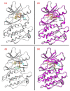
(Panel (A)) Ligand-based homology model for the T599dup B-RAF variant complexed with Dabrafenib (from PDB: 4XV2, colored in gold). The T599 and the newly inserted T600 residues are colored orange and labeled in black and orange, respectively. (Panel (B)) Superposition of the backbones of the T599dup B-RAF/Dabrafenib ligand-based homology model with the reference crystal structure of B-RAF V600E (PDB code: 6P7G). The E600 residue of the V600E variant is colored magenta and labeled in red. The RMSD of the backbones of the two superposed structures was 0.60 Å. (Panel (C)) Ligand-based homology model for the A598T599_insV B-RAF variant complexed with Dabrafenib. The A598 and the T600 residues are colored orange and labeled in black; the newly inserted V599 residue is colored and labeled in green. (Panel (D)) Superposition of the backbones of the A598_T599_insV B-RAF/Dabrafenib ligand-based homology model with the reference crystal structure of B-RAF V600E. The RMSD of the backbones of the two superposed structures was 0.96 Å. The hydrogen bonds are highlighted with cyan-colored sticks, whose thickness is proportional to their strength. All the images represented in the panels were created and rendered with MOE.
Figure 5
Figure 5
(Panel (A)) Dabrafenib (colored in gold) in the binding pocket of the T599dup ligand-based homology model. The T599 and the newly inserted T600 residues are colored in orange and labeled in black and orange, respectively. (Panel (B)) Superposition of the T599dup B-RAF/Dabrafenib ligand-based homology model with the best docking pose obtained with Glide (colored in dark green). The RMSD between the two Dabrafenib poses was 0.22 Å. (Panel (C)) Dabrafenib in the binding pocket of the A598_T599_insV ligand-based homology model. The A598 and the T600 residues are colored orange and labeled in black; the newly inserted V599 residue is colored and labeled in green. (Panel (D)) Superposition of the A598_T599_insV B-RAF/Dabrafenib ligand-based homology model with the best docking pose obtained with Glide. The RMSD between the two Dabrafenib poses was 0.38 Å. The hydrogen bonds are highlighted with cyan-colored sticks, whose thickness is proportional to their strength. All the images represented in the panels were created and rendered with MOE.

References

    1. Cancer Genome Atlas Network Genomic Classification of Cutaneous Melanoma. Cell. 2015;161:1681–1696. doi: 10.1016/j.cell.2015.05.044. - DOI - PMC - PubMed
    1. Menzer C., Menzies A.M., Carlino M.S., Reijers I., Groen E.J., Eigentler T., de Groot J.W.B., van der Veldt A.A.M., Johnson D.B., Meiss F., et al. Targeted Therapy in Advanced Melanoma with Rare BRAF Mutations. J. Clin. Oncol. 2019;37:3142–3151. doi: 10.1200/JCO.19.00489. - DOI - PMC - PubMed
    1. Johnson D.B., Zhao F., Noel M., Riely G.J., Mitchell E.P., Wright J.J., Chen H.X., Gray R.J., Li S., McShane L.M., et al. Trametinib Activity in Patients with Solid Tumors and Lymphomas Harboring BRAF Non-V600 Mutations or Fusions: Results from NCI-MATCH (EAY131) Clin. Cancer Res. 2020;26:1812–1819. doi: 10.1158/1078-0432.CCR-19-3443. - DOI - PMC - PubMed
    1. Bahadoran P., Allegra M., Le Duff F., Long-Mira E., Hofman P., Giacchero D., Passeron T., Lacour J.-P., Ballotti R. Major Clinical Response to a BRAF Inhibitor in a Patient with a BRAF L597R-Mutated Melanoma. J. Clin. Oncol. 2013;31:e324–e326. doi: 10.1200/JCO.2012.46.1061. - DOI - PubMed
    1. Zhang S., Yang Z., Cheng Y., Guo X., Liu C., Wang S., Zhang L. BRAF L485-P490 Deletion Mutant Metastatic Melanoma Sensitive to BRAF and MEK Inhibition: A Case Report and Literature Review. Front. Pharmacol. 2022;13:1019217. doi: 10.3389/fphar.2022.1019217. - DOI - PMC - PubMed