

Sortilin Is Upregulated in Osteoarthritis-Dependent Cartilage Calcification and Associated with Cellular Senescence
- PMID: 37569721
- PMCID: PMC10418692
- DOI: 10.3390/ijms241512343
Sortilin Is Upregulated in Osteoarthritis-Dependent Cartilage Calcification and Associated with Cellular Senescence
Abstract
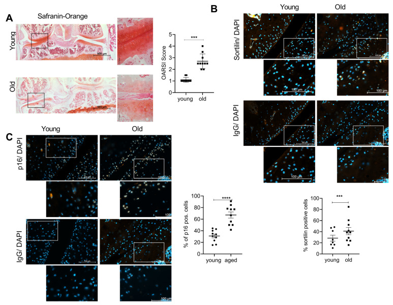
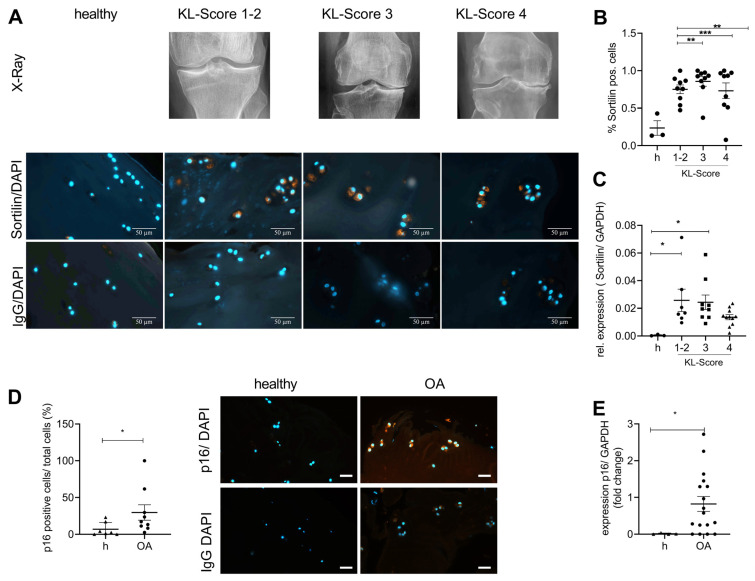
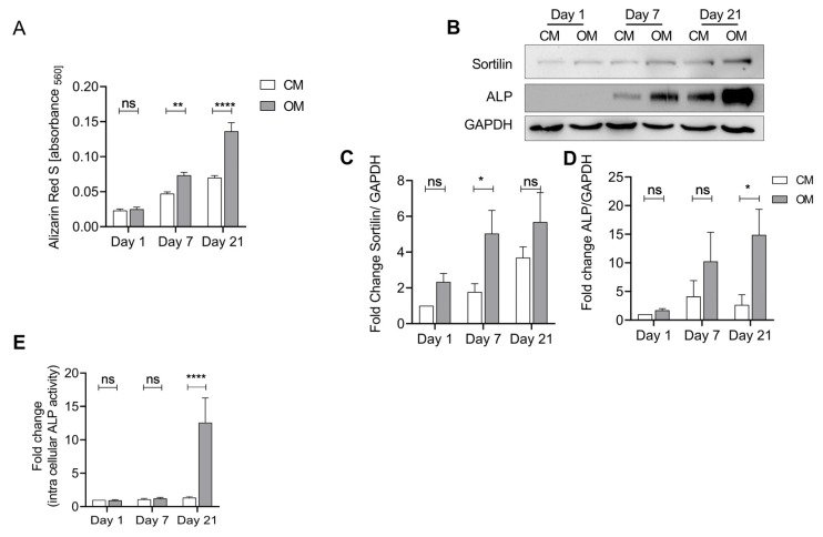
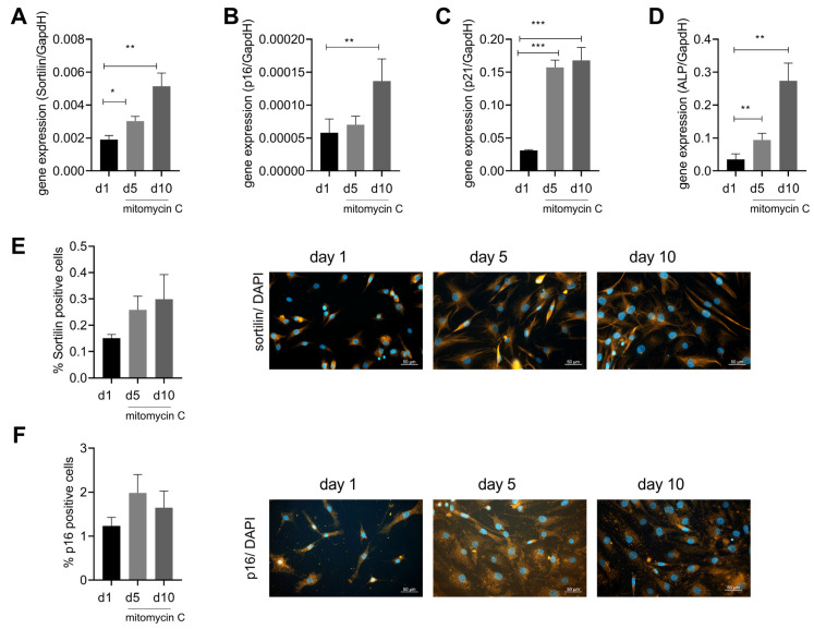
Osteoarthritis (OA) is a chronic joint disease characterized by articular cartilage calcification, loss of articular cartilage, bone changes, pain, and disability. Cartilage calcification is one hallmark of OA and is predominantly caused by basic calcium crystals formed due to an imbalance of the pyrophosphate pathway. Sortilin is a transmembrane protein that contributes to vascular calcification in atherosclerosis by externalizing alkaline phosphatase (ALP)-containing vesicles. Calcification in atherosclerosis and osteoarthritis has been associated with cellular senescence. The aim of this study was to investigate the potential role of sortilin and senescence in osteoarthritis-dependent cartilage calcification. Osteoarthritic cartilage from human knee joints was collected after joint replacement, and samples were analyzed by immunohistochemistry and quantitative RT-PCR analysis. Human chondrocytes were treated with osteogenic medium for up to 21 days to induce calcification. Western blots for sortilin and ALP, as well as an ALP activity assay, were performed. Human chondrocytes were treated with mitomycin C to induce senescence, and sortilin expression was quantified at the protein and gene levels. Sections of knee joints from a murine model of osteoarthritis were stained for sortilin and p16 and analyzed by immunohistochemistry. Treatment of wild-type chondrocytes using an osteogenic medium similar to human chondrocytes was performed. Osteoarthritic cartilage from mouse and human knee joints showed an increased number of sortilin and p16-positive chondrocytes compared to healthy cartilage. This observation was corroborated by increased gene expression of sortilin and p16 in mild and moderate osteoarthritic cartilage samples. To investigate the mechanism of sortilin regulation, human chondrocytes were treated with osteogenic medium to induce calcification. Sortilin protein levels and expression were increased after 7 days of stimulation, whereas ALP levels and activity were upregulated after 21 days of stimulation. Similar observations were made in a murine osteoarthritis model. Mechanistically, senescent chondrocytes induced by mitomycin C showed an upregulation of sortilin and ALP gene expression compared to non-senescent chondrocytes. Our data indicate that sortilin and ALP are upregulated during cartilage calcification, which is associated with chondrocyte senescence and thus might contribute to the pathogenesis of osteoarthritis. Cellular senescence seems to induce sortilin expression.
Keywords: calcification; cartilage; chondrocytes; osteoarthritis; senescence; sortilin.
Conflict of interest statement
The authors declare that the research was conducted in the absence of any commercial or financial relationships that could be construed as a potential conflict of interest.
Figures
Similar articles
-
Chondrocytes From Osteoarthritic and Chondrocalcinosis Cartilage Represent Different Phenotypes.Front Cell Dev Biol. 2021 Apr 26;9:622287. doi: 10.3389/fcell.2021.622287. eCollection 2021. Front Cell Dev Biol. 2021. PMID: 33981699 Free PMC article.
-
Endothelin-1 induces chondrocyte senescence and cartilage damage via endothelin receptor type B in a post-traumatic osteoarthritis mouse model.Osteoarthritis Cartilage. 2020 Dec;28(12):1559-1571. doi: 10.1016/j.joca.2020.08.006. Epub 2020 Aug 25. Osteoarthritis Cartilage. 2020. PMID: 32858189
-
Adenosine A2A receptor activation reduces chondrocyte senescence.FASEB J. 2023 Apr;37(4):e22838. doi: 10.1096/fj.202201212RR. FASEB J. 2023. PMID: 36884388 Free PMC article.
-
Cellular senescence in knee osteoarthritis: molecular mechanisms and therapeutic implications.Ageing Res Rev. 2021 Sep;70:101413. doi: 10.1016/j.arr.2021.101413. Epub 2021 Jul 21. Ageing Res Rev. 2021. PMID: 34298194 Review.
-
Cartilage calcification in osteoarthritis: mechanisms and clinical relevance.Nat Rev Rheumatol. 2023 Jan;19(1):10-27. doi: 10.1038/s41584-022-00875-4. Epub 2022 Dec 12. Nat Rev Rheumatol. 2023. PMID: 36509917 Review.
Cited by
-
CPP-calcification of articular cartilage is associated with elevated cytokine levels in synovial fluid.Front Cell Dev Biol. 2025 Mar 19;13:1535530. doi: 10.3389/fcell.2025.1535530. eCollection 2025. Front Cell Dev Biol. 2025. PMID: 40177128 Free PMC article.
-
Sortilin and its potential role in cardiovascular pathology.Egypt Heart J. 2024 Jun 24;76(1):78. doi: 10.1186/s43044-024-00512-3. Egypt Heart J. 2024. PMID: 38913092 Free PMC article. Review.
-
Circulating Proneurotensin Levels Predict Impaired Bone Mineralisation in Postmenopausal Women With Type 2 Diabetes Mellitus.Diabetes Metab Res Rev. 2025 Jan;41(1):e70018. doi: 10.1002/dmrr.70018. Diabetes Metab Res Rev. 2025. PMID: 39655684 Free PMC article.
References
Grants and funding
LinkOut - more resources
Full Text Sources
