. 2023 Aug 2;24(15):12367.
doi: 10.3390/ijms241512367.

Coffea arabica Extract Attenuates Atopic Dermatitis-like Skin Lesions by Regulating NLRP3 Inflammasome Expression and Skin Barrier Functions

Affiliations

Coffea arabica Extract Attenuates Atopic Dermatitis-like Skin Lesions by Regulating NLRP3 Inflammasome Expression and Skin Barrier Functions

Qiao-Xin Chang et al. Int J Mol Sci. .

Abstract

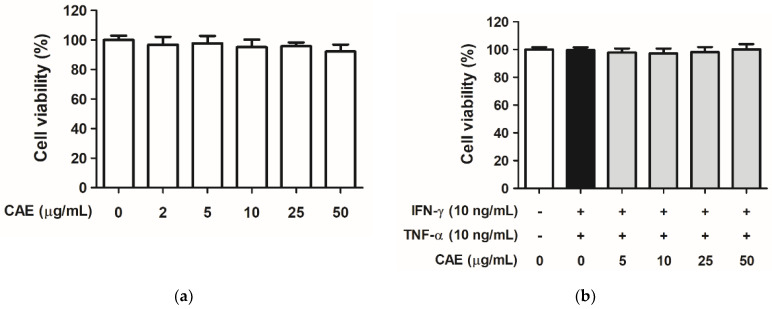
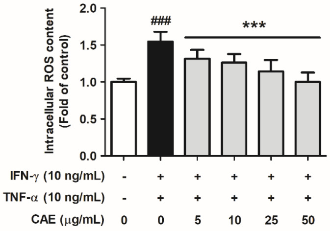
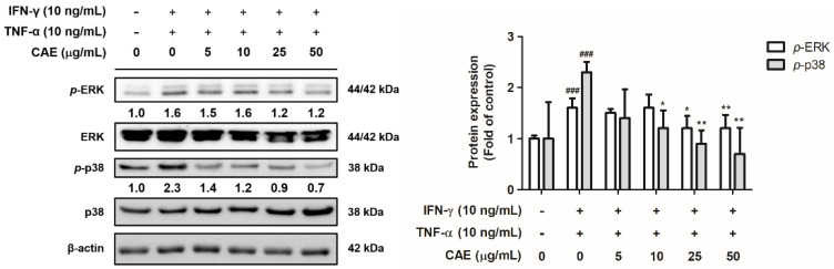
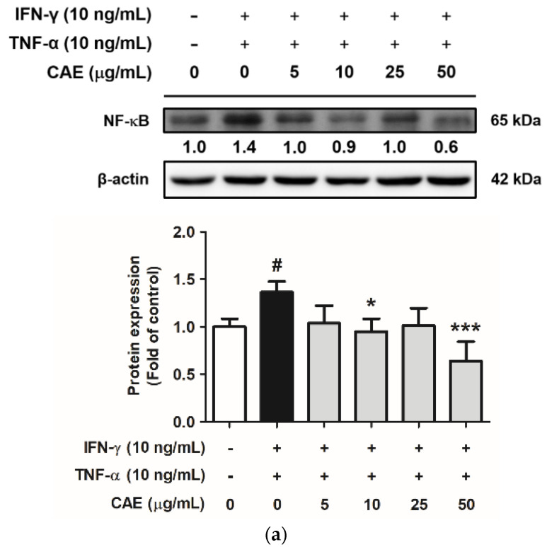
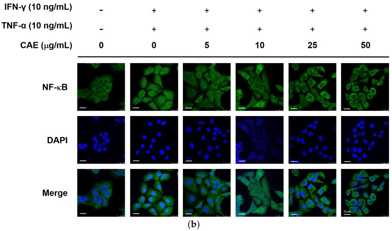
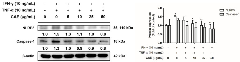
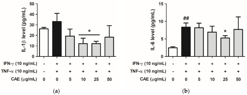
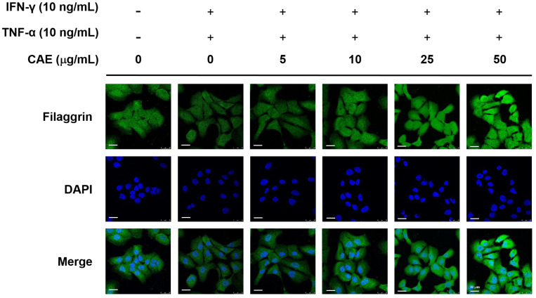
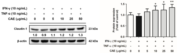
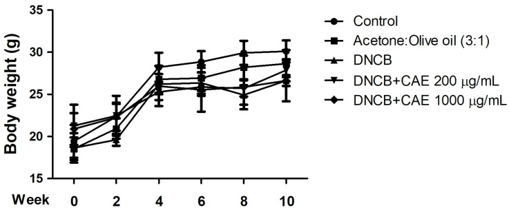
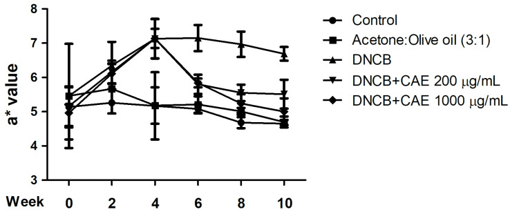
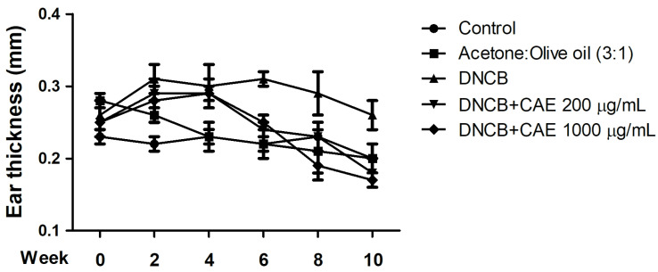
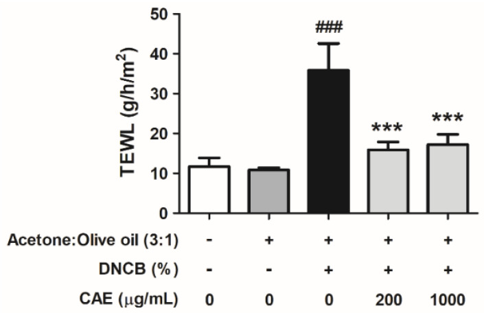
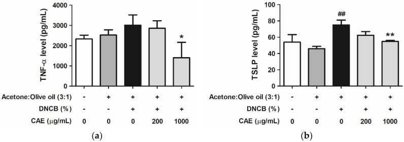
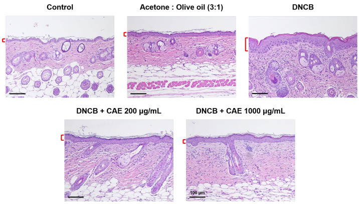
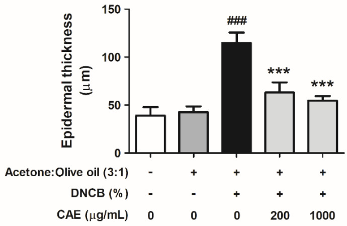
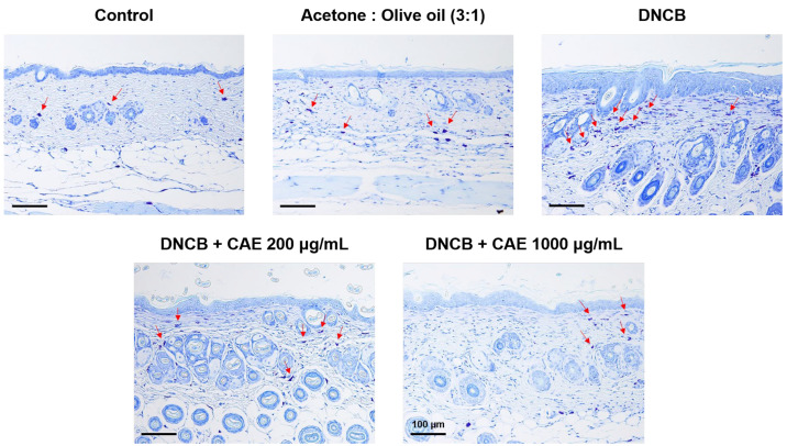
Atopic dermatitis (AD) is a common skin disease worldwide. The major causes of AD are skin barrier defects, immune dysfunction, and oxidative stress. In this study, we investigated the anti-oxidation and anti-inflammation effects of Coffea arabica extract (CAE) and its regulation of the skin barrier and immune functions in AD. In vitro experiments revealed that CAE decreased the reactive oxygen species levels and inhibited the translocation of nuclear factor-κB (NF-κB), further reducing the secretion of interleukin (IL)-1β and IL-6 induced by interferon-γ (IFN-γ)/tumor necrosis factor-α (TNF-α). Moreover, CAE decreased IFN-γ/TNF-α-induced NLR family pyrin domain-containing 3 (NLRP3), caspase-1, high-mobility group box 1 (HMGB1), and receptor for advanced glycation end products (RAGE) expression levels. It also restored the protein levels of skin barrier function-related markers including filaggrin and claudin-1. In vivo experiments revealed that CAE not only reduced the redness of the backs of mice caused by 2,4-dinitrochlorobenzene (DNCB) but also reduced the levels of pro-inflammatory factors in their skin. CAE also reduced transepidermal water loss (TEWL) and immune cell infiltration in DNCB-treated mice. Overall, CAE exerted anti-oxidation and anti-inflammation effects and ameliorated skin barrier dysfunction, suggesting its potential as an active ingredient for AD treatment.

Keywords: Coffea arabica; anti-inflammation; anti-oxidation; atopic dermatitis; immune; inflammasome; skin barrier function.


Conflict of interest statement

The authors declare no conflict of interest.

Figures

Figure 1
Figure 1
Cell viabilities of human skin keratinocytes after (a) C. arabica extract (CAE) treatment and (b) CAE treatment after IFN-γ and TNF-α induction for 24 h. CAE did not exhibit cytotoxic effects on HaCaT cells.
Figure 2
Figure 2
Effects of CAE on intracellular ROS levels in IFN-γ- and TNF-α-induced human skin keratinocytes. ###, p < 0.001 vs. non-induced group. ***, p < 0.001 vs. IFN-γ/TNF-α-induced group. CAE significantly attenuated IFN-γ- and TNF-α-induced oxidative stress in HaCaT cells.
Figure 3
Figure 3
Effects of CAE on p-ERK and p-p38 protein expression levels in IFN-γ- and TNF-α-induced human skin keratinocytes after 24 h. ###, p < 0.001 vs. non-induced group. *, p < 0.05 and **, p < 0.01 vs. IFN-γ/TNF-α-induced group. CAE significantly reduced the IFN-γ- and TNF-α-induced p-ERK and p-p38 expression in HaCaT cells.
Figure 4
Figure 4
Effects of CAE on (a) protein expression levels and (b) nuclear translocation of NF-κB in IFN-γ- and TNF-α-induced human skin keratinocytes after 24 h. Immunofluorescence staining of NF-κB (green) and nucleus (DAPI, blue) (scale bar = 20 µm). #, p < 0.05 vs. non-induced group. *, p < 0.05 and ***, p < 0.001 vs. IFN-γ/TNF-α-induced group. CAE decreased the expression and the translocation of NF-κB stimulated by IFN-γ and TNF-α in HaCaT cells.
Figure 4
Figure 4
Effects of CAE on (a) protein expression levels and (b) nuclear translocation of NF-κB in IFN-γ- and TNF-α-induced human skin keratinocytes after 24 h. Immunofluorescence staining of NF-κB (green) and nucleus (DAPI, blue) (scale bar = 20 µm). #, p < 0.05 vs. non-induced group. *, p < 0.05 and ***, p < 0.001 vs. IFN-γ/TNF-α-induced group. CAE decreased the expression and the translocation of NF-κB stimulated by IFN-γ and TNF-α in HaCaT cells.
Figure 5
Figure 5
Effects of CAE on the NLR family pyrin domain-containing 3 (NLRP3) and caspase-1 protein expression levels in IFN-γ- and TNF-α-induced human skin keratinocytes after 24 h. #, p < 0.05 vs. non-induced group. *, p < 0.05 and **, p < 0.01 vs. IFN-γ/TNF-α-induced group. CAE decreased the expression of NLRP3 and caspase-1 stimulated by IFN-γ and TNF-α in HaCaT cells.
Figure 6
Figure 6
Effects of CAE on the secretion of (a) interleukin (IL)-1β and (b) IL-6 in IFN-γ- and TNF-α-induced human skin keratinocytes after 24 h. ##, p < 0.01 vs. non-induced group. *, p < 0.05 vs. IFN-γ/TNF-α-induced group. CAE decreased the IFN-γ- and TNF-α-induced IL-1β and IL-6 levels in HaCaT cells.
Figure 7
Figure 7
Effects of CAE on HMGB1 and RAGE protein expression levels in IFN-γ- and TNF-α-induced human skin keratinocytes after 24 h. *, p < 0.05, **, p < 0.01 and ***, p < 0.001 vs. IFN-γ/TNF-α-induced group. CAE significantly reduced the IFN-γ- and TNF-α-induced HMGB1 and RAGE expression in HaCaT cells.
Figure 8
Figure 8
Effects of CAE on the nuclear translocation of filaggrin in IFN-γ- and TNF-α-induced human skin keratinocytes after 24 h. Immunofluorescence staining of filaggrin (green) and nucleus (DAPI, blue) (scale bar = 20 µm). CAE increased the translocation of filaggrin stimulated by IFN-γ and TNF-α in HaCaT cells.
Figure 9
Figure 9
Effects of CAE on claudin-1 protein expression levels in IFN-γ- and TNF-α-induced human skin keratinocytes after 24 h. *, p < 0.05 and **, p < 0.01 vs. IFN-γ/TNF-α-induced group. CAE increased the expression of claudin-1 stimulated by IFN-γ and TNF-α in HaCaT cells.
Figure 10
Figure 10
Body weights of BALB/c mice during 10 weeks of treatment. Non-significant difference between the groups.
Figure 11
Figure 11
Effects of CAE inflammation indicated by a* values in DNCB-induced BALB/c mice during 10 weeks of treatment. CAE improved DNCB-induced erythema and inflammation in AD.
Figure 12
Figure 12
Effects of CAE on ear thickness in DNCB-induced BALB/c mice during 10 weeks of treatment. CAE inhibited DNCB-induced skin edema.
Figure 13
Figure 13
Effects of CAE on TEWL in DNCB-induced BALB/c mice in the 10th week of treatment. ###, p < 0.001 vs. control group. ***, p < 0.001 vs. DNCB-induced group. CAE ameliorated DNCB-induced skin barrier dysfunction.
Figure 14
Figure 14
Effects of CAE on the secretion of (a) TNF-α and (b) TSLP in DNCB-induced BALB/c mice in the 10th week of treatment. ##, p < 0.01 vs. control group. *, p < 0.05 and **, p < 0.01 vs. DNCB-induced group. CAE decreased DNCB-induced TNF-α and TSLP levels.
Figure 15
Figure 15
Histopathological characterization of H&E-stained BALB/c mice treated with DNCB and CAE (scale bar = 100 µm). The red mark represents the thickness of the skin epidermis CAE decreased DNCB-induced skin epidermal thickness.
Figure 16
Figure 16
Effects of CAE on the epidermal thickness of H&E-stained DNCB-induced BALB/c mice in the 10th week of treatment. ###, p < 0.001 vs. control group. ***, p < 0.001 vs. DNCB-induced group. CAE significantly decreased DNCB-induced epidermal thickness.
Figure 17
Figure 17
Histopathological characterization (indicated by arrows) of toluidine blue-stained BALB/c mice treated with DNCB and CAE (scale bar = 100 µm). CAE attenuated DNCB-induced AD-like skin lesions.

Similar articles

Cited by

References

    1. Bylund S., von Kobyletzki L.B., Svalstedt M., Svensson Å. Prevalence and incidence of atopic dermatitis: A systematic review. Acta Derm.-Venereol. 2020;100:320–329. doi: 10.2340/00015555-3510. - DOI - PMC - PubMed
    1. Nguyen M., Pona A., Kolli S.S., Feldman S.R., Strowd L.C. Recent insights in atopic dermatitis pathogenesis, treatment, and disease impact. J. Dermatol. Dermatol. Surg. 2019;23:66–72.
    1. Chiricozzi A., Maurelli M., Calabrese L., Peris K., Girolomoni G. Overview of Atopic Dermatitis in Different Ethnic Groups. J. Clin. Med. 2023;12:2701. doi: 10.3390/jcm12072701. - DOI - PMC - PubMed
    1. Tollefson M.M., Bruckner A.L. Atopic dermatitis: Skin-directed management. Pediatrics. 2014;134:e1735–e1744. doi: 10.1542/peds.2014-2812. - DOI - PubMed
    1. Elias P.M., Steinhoff M. “Outside-to-inside”(and now back to “outside”) pathogenic mechanisms in atopic dermatitis. J. Investig. Dermatol. 2008;128:1067–1070. doi: 10.1038/jid.2008.88. - DOI - PMC - PubMed

LinkOut - more resources