Review
. 2023 Aug 3;24(15):12390.
doi: 10.3390/ijms241512390.

Asbestos and Iron

Affiliations
Review

Asbestos and Iron

Andrew J Ghio et al. Int J Mol Sci. .

Abstract

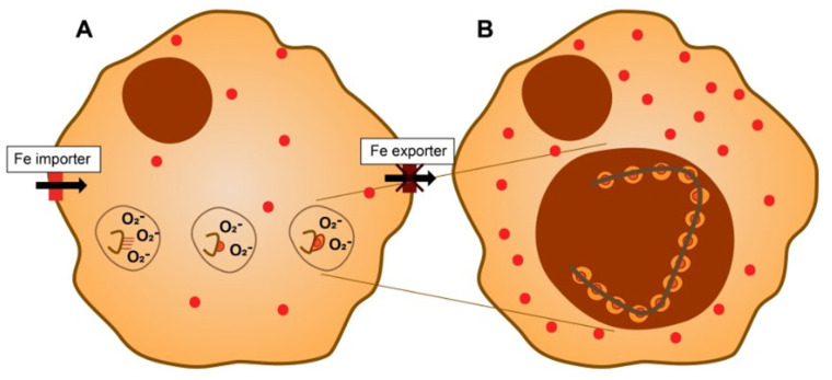
Theories of disease pathogenesis following asbestos exposure have focused on the participation of iron. After exposure, an open network of negatively charged functional groups on the fiber surface complexes host metals with a preference for iron. Competition for iron between the host and the asbestos results in a functional metal deficiency. The homeostasis of iron in the host is modified by the cell response, including increased import to correct the loss of the metal to the fiber surface. The biological effects of asbestos develop in response to and are associated with the disruption of iron homeostasis. Cell iron deficiency in the host following fiber exposure activates kinases and transcription factors, which are associated with the release of mediators coordinating both inflammatory and fibrotic responses. Relative to serpentine chrysotile, the clearance of amphiboles is incomplete, resulting in translocation to the mesothelial surface of the pleura. Since the biological effect of asbestos is dependent on retention of the fiber, the sequestration of iron by the surface, and functional iron deficiency in the cell, the greater clearance (i.e., decreased persistence) of chrysotile results in its diminished impact. An inability to clear asbestos from the lower respiratory tract initiates a host process of iron biomineralization (i.e., asbestos body formation). Host cells attempt to mobilize the metal sequestered by the fiber surface by producing superoxide at the phagosome membrane. The subsequent ferrous cation is oxidized and undergoes hydrolysis, creating poorly crystalline iron oxyhydroxide (i.e., ferrihydrite) included in the coat of the asbestos body.

Keywords: alveolar macrophages; asbestos; ferritin; iron; lung diseases.


Conflict of interest statement

The authors declare no conflict of interest.

Figures

Figure 1
Figure 1
Clearance of asbestos from the lower respiratory tract. Exposure to longer amphibole fibers can result in “frustrated phagocytosis” and failure of clearance pathways, while shorter fibers and fragments can be successfully phagocytosed and removed via the mucociliary pathway. Phagosomal degradation of amphibole with release of metals does not occur (A). In contrast, longer chrysotile fibers are phagocytosed and the brucite layer is destroyed in an acidic environment with release of metal (e.g., magnesium) constituents. This results in fragments that can eliminated from the lower respiratory tract via the mucociliary clearance pathway (B).
Figure 2
Figure 2
Cell exposure to asbestos and iron homeostasis. Functional groups (e.g., -Si-O groups) on the surface of the fibrous silica will complex host sources of intracellular iron. There is a loss of metal required for critical functions in the cell. Mitochondria are especially vulnerable as processes in this organelle demonstrate significant dependence on the availability of this metal (e.g., Krebs cycle and electron transport system).
Figure 3
Figure 3
Exposure to asbestos causes a functional iron deficiency. The host response reflects an attempt to return to normal iron homeostasis with metal available for critical functions and cell survival. This includes inflammation and fibrosis.
Figure 4
Figure 4
The biological effects of asbestos and iron. Decreased iron availability affects kinase and transcription factor activation, which coordinate a release of mediators relevant to inflammatory and fibrotic responses. An influx of inflammatory cells (e.g., macrophages and neutrophils) corresponds with the decreased metal availability. There is also an increased number of fibroblasts and a deposition of collagen (represented by blue helical units), elastin (represented by yellow units), and extracellular polymeric substances, which correspond to functional iron deficiency.
Figure 5
Figure 5
Asbestos body development and iron. In the phagolysosome, asbestos complexes cell iron, impacting functional iron deficiency (A). The host cell responds with increases in the import and decreases in the export of iron, elevating storage of the metal in ferritin (represented by red dots). In addition, NADPH oxidoreductases at the phagosome membrane will increase the generation of superoxide to reduce Fe3+ to Fe2+, which is released by surface functional groups. The Fe2+ reacts with oxygen to form iron to form oxyhydroxides including ferrihydrite. Nanoparticulate iron oxyhydroxides provide a large surface, which can react with additional metals (e.g., iron, calcium, magnesium, gadolinium, and radium). The iron-containing structure that results corresponds to the template provided by the position of the functional groups (homogeneous, segmental, barbell, etc.) at the asbestos surface (B).

References

    1. Cooke W.E. Fibrosis of the lungs due to the inhalation of asbestos dust. Br. Med. J. 1924;2:147. doi: 10.1136/bmj.2.3317.147. - DOI - PMC - PubMed
    1. Breslow L. Industrial aspects of bronchiogenic neoplasms. Dis. Chest. 1955;28:421–430. doi: 10.1378/chest.28.4.421. - DOI - PubMed
    1. Doll R. Mortality from lung cancer in asbestos workers. Br. J. Ind. Med. 1955;12:81–86. doi: 10.1136/oem.12.2.81. - DOI - PMC - PubMed
    1. Wagner J.C., Sleggs C.A., Marchand P. Diffuse pleural mesothelioma and asbestos exposure in the North Western Cape Province. Br. J. Ind. Med. 1960;17:260–271. doi: 10.1136/oem.17.4.260. - DOI - PMC - PubMed
    1. Nagai H., Okazaki Y., Chew S.H., Misawa N., Yasui H., Toyokuni S. Deferasirox induces mesenchymal-epithelial transition in crocidolite-induced mesothelial carcinogenesis in rats. Cancer Prev. Res. 2013;6:1222–1230. doi: 10.1158/1940-6207.CAPR-13-0244. - DOI - PubMed

LinkOut - more resources