. 2023 Aug 4;24(15):12424.
doi: 10.3390/ijms241512424.

Cannabidiol and Cannabigerol Modify the Composition and Physicochemical Properties of Keratinocyte Membranes Exposed to UVA

Affiliations

Cannabidiol and Cannabigerol Modify the Composition and Physicochemical Properties of Keratinocyte Membranes Exposed to UVA

Adam Wroński et al. Int J Mol Sci. .

Abstract

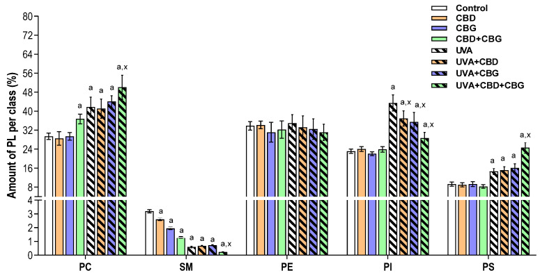
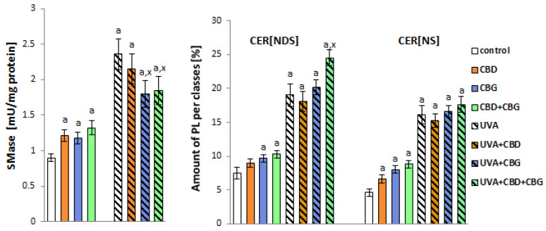
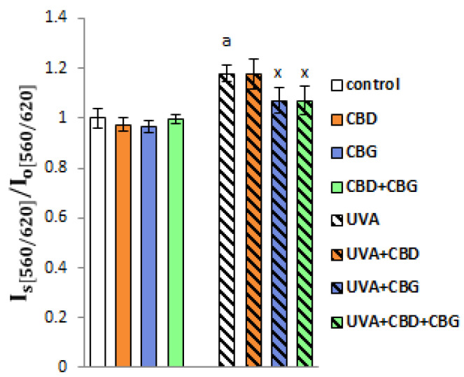
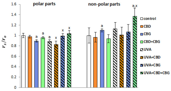
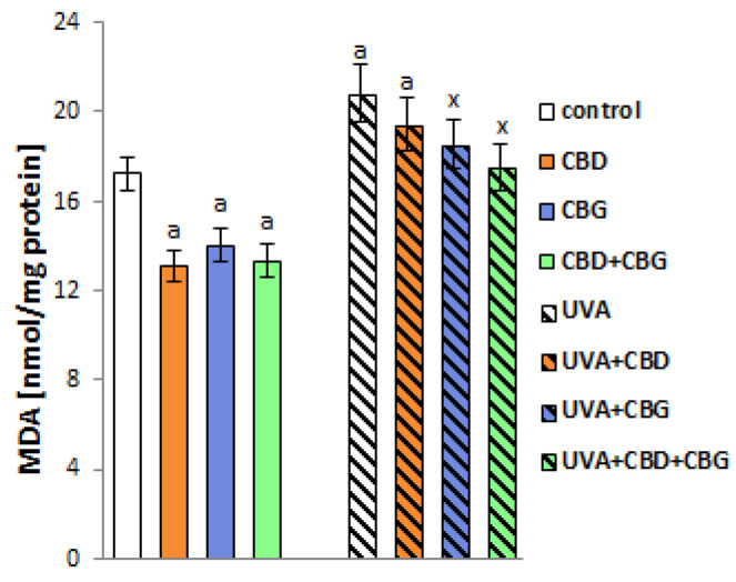
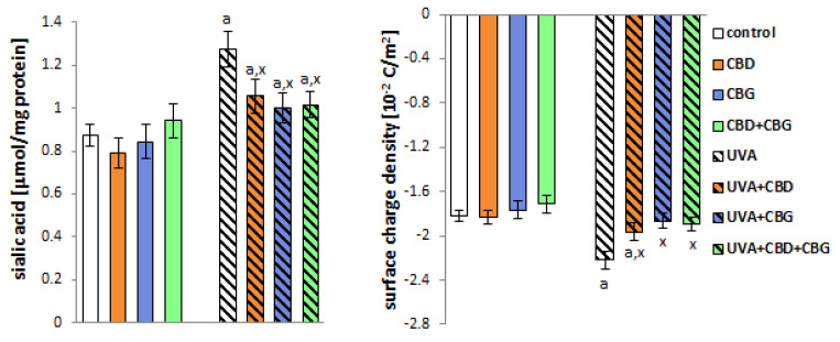
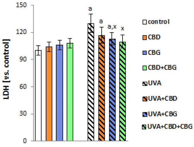
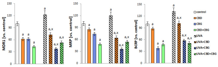
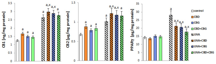
The action of UVA radiation (both that derived from solar radiation and that used in the treatment of skin diseases) modifies the function and composition of keratinocyte membranes. Therefore, this study aimed to assess the effects of phytocannabinoids (CBD and CBG), used singly and in combination, on the contents of phospholipids, ceramides, lipid rafts and sialic acid in keratinocyte membranes exposed to UVA radiation, together with their structure and functionality. The phytocannabinoids, especially in combination (CBD+CBG), partially prevented increased levels of phosphatidylinositols and sialic acid from occurring and sphingomyelinase activity after the UVA exposure of keratinocytes. This was accompanied by a reduction in the formation of lipid rafts and malondialdehyde, which correlated with the parameters responsible for the integrity and functionality of the keratinocyte membrane (membrane fluidity and permeability and the activity of transmembrane transporters), compared to UVA-irradiated cells. This suggests that the simultaneous use of two phytocannabinoids may have a protective effect on healthy cells, without significantly reducing the therapeutic effect of UV radiation used to treat skin diseases such as psoriasis.

Keywords: ceramides; lipid rafts; membrane electrical charge; membrane fluidity; phospholipids; phytocannabinoids; transmembrane transporters.


Conflict of interest statement

The authors declare no conflict of interest.

Figures

Figure 1
Figure 1
Relative phospholipid classes in the following keratinocyte groups: control; cultured with CBD (5 μM) for 24 h; cultured with CBG (1 μM) for 24 h; cultured with CBD (5 μM) and CBG (1 μM) for 24 h; irradiated with UVA (30 J/cm2); irradiated with UVA (30 J/cm2) and cultured with CBD (5 μM) for 24 h after irradiation; irradiated with UVA (30 J/cm2) and cultured with CBG (1 μM) for 24 h; irradiated with UVA (30 J/cm2) and cultured with CBD (5 μM) and CBG (1 μM) for 24 h. Values are mean ± SD, p < 0.05; a, significantly different from control; x, significantly different from UVA group.
Figure 2
Figure 2
SMase activity panel and relative ceramide content within CER[NDS] and CER[NS] classes in the following keratinocyte groups: control; cultured with CBD (5 μM) for 24 h; cultured with CBG (1 μM) for 24 h; cultured with CBD (5 μM) and CBG (1 μM) for 24 h; irradiated with UVA (30 J/cm2); irradiated with UVA (30 J/cm2) and cultured with CBD (5 μM) for 24 h after irradiation; irradiated with UVA (30 J/cm2) and cultured with CBG (1 μM) for 24 h; irradiated with UVA (30 J/cm2) and cultured with CBD (5 μM) and CBG (1 μM) for 24 h. Values are mean ± SD, p < 0.05; a, significantly different from control; x, significantly different from UVA group.
Figure 3
Figure 3
Formation of lipid rafts in the following keratinocyte groups: control; cultured with CBD (5 μM) for 24 h; cultured with CBG (1 μM) for 24 h; cultured with CBD (5 μM) and CBG (1 μM) for 24 h; irradiated with UVA (30 J/cm2); irradiated with UVA (30 J/cm2) and cultured with CBD (5 μM) for 24 h after irradiation; irradiated with UVA (30 J/cm2) and cultured with CBG (1 μM) for 24 h; irradiated with UVA (30 J/cm2) and cultured with CBD (5 μM) and CBG (1 μM) for 24 h. Values are mean ± SD, p < 0.05; a, significantly different from control; x, significantly different from UVA group. Is is the ratio λem.1/λem.2 for samples, while I0 is the ratio λem.1/λem.2 for the control cells.
Figure 4
Figure 4
Fluorescence anisotropy of membrane phospholipids in their polar (TMA-DPH staining) and non-polar (DPH staining) parts in the following keratinocyte groups: control; cultured with CBD (5 μM) for 24 h; cultured with CBG (1 μM) for 24 h; cultured with CBD (5 μM) and CBG (1 μM) for 24 h; irradiated with UVA (30 J/cm2); irradiated with UVA (30 J/cm2) and cultured with CBD (5 μM) for 24 h after irradiation; irradiated with UVA (30 J/cm2) and cultured with CBG (1 μM) for 24 h; irradiated with UVA (30 J/cm2) and cultured with CBD (5 μM) and CBG (1 μM) for 24 h. Values are mean ± SD, p < 0.05; a, significantly different from control; x, significantly different from UVA group. The results are presented as the rs/r0 ratio, where rs and r0 are the anisotropy values for the test and control samples, respectively.
Figure 5
Figure 5
The levels of lipid peroxidation product (MDA) in the following keratinocyte groups: control; cultured with CBD (5 μM) for 24 h; cultured with CBG (1 μM) for 24 h; cultured with CBD (5 μM) and CBG (1 μM) for 24 h; irradiated with UVA (30 J/cm2); irradiated with UVA (30 J/cm2) and cultured with CBD (5 μM) for 24 h after irradiation; irradiated with UVA (30 J/cm2) and cultured with CBG (1 μM) for 24 h; irradiated with UVA (30 J/cm2) and cultured with CBD (5 μM) and CBG (1 μM) for 24 h. Values are mean ± SD, p < 0.05; a, significantly different from control; x, significantly different from UVA group.
Figure 6
Figure 6
The sialic acid content and the surface charge density of the following keratinocyte groups: control; cultured with CBD (5 μM) for 24 h; cultured with CBG (1 μM) for 24 h; cultured with CBD (5 μM) and CBG (1 μM) for 24 h; irradiated with UVA (30 J/cm2); irradiated with UVA (30 J/cm2) and cultured with CBD (5 μM) for 24 h after irradiation; irradiated with UVA (30 J/cm2) and cultured with CBG (1 μM) for 24 h; irradiated with UVA (30 J/cm2) and cultured with CBD (5 μM) and CBG (1 μM) for 24 h. Values are mean ± SD, p < 0.05; a, significantly different from control; x, significantly different from UVA group.
Figure 7
Figure 7
The activity of lactate dehydrogenase (LDH) in the media of the following keratinocyte groups: control; cultured with CBD (5 μM) for 24 h; cultured with CBG (1 μM) for 24 h; cultured with CBD (5 μM) and CBG (1 μM) for 24 h; irradiated with UVA (30 J/cm2); irradiated with UVA (30 J/cm2) and cultured with CBD (5 μM) for 24 h after irradiation; irradiated with UVA (30 J/cm2) and cultured with CBG (1 μM) for 24 h; irradiated with UVA (30 J/cm2) and cultured with CBD (5 μM) and CBG (1 μM) for 24 h. Values are mean ± SD, p < 0.05; a, significantly different from control; x, significantly different from UVA group.
Figure 8
Figure 8
The activity of ABC-cassette transporters (MDR1, MRP, BCRP) in the following keratinocyte groups: control; cultured with CBD (5 μM) for 24 h; cultured with CBG (1 μM) for 24 h; cultured with CBD (5 μM) and CBG (1 μM) for 24 h; irradiated with UVA (30 J/cm2); irradiated with UVA (30 J/cm2) and cultured with CBD (5 μM) for 24 h after irradiation; irradiated with UVA (30 J/cm2) and cultured with CBG (1 μM) for 24 h; irradiated with UVA (30 J/cm2) and cultured with CBD (5 μM) and CBG (1 μM) for 24 h. Values are mean ± SD, p < 0.05; a, significantly different from control; x, significantly different from UVA group.
Figure 9
Figure 9
The expression of receptors (CB1, CB2, PPARγ) in the following keratinocyte groups: control; cultured with CBD (5 μM) for 24 h; cultured with CBG (1 μM) for 24 h; cultured with CBD (5 μM) and CBG (1 μM) for 24 h; irradiated with UVA (30 J/cm2 ); irradiated with UVA (30 J/cm2) and cultured with CBD (5 μM) for 24 h after irradiation; irradiated with UVA (30 J/cm2) and cultured with CBG (1 μM) for 24 h; irradiated with UVA (30 J/cm2) and cultured with CBD (5 μM) and CBG (1 μM) for 24 h. Values are mean ± SD, p < 0.05; a, significantly different from control; x, significantly different from UVA group.

Similar articles

Cited by

References

    1. Gilaberte Y., Prieto-Torres L., Pastushenko I., Juarranz Á. Anatomy and Function of the Skin. In: Hamblin M.R., Avci P., Prow T.W., editors. Nanoscience in Dermatology. Academic Press; Cambridge, MA, USA: 2016. pp. 1–14. Chapter 1.
    1. Nicolson G.L., Ferreira de Mattos G. A brief introduction to some aspects of the fluid–mosaic model of cell membrane structure and its importance in membrane lipid replacement. Membranes. 2021;11:947. doi: 10.3390/membranes11120947. - DOI - PMC - PubMed
    1. Sezgin E., Levental I., Mayor S., Eggeling C. The mystery of membrane organization: Composition, regulation and roles of lipid rafts. Nat. Rev. Mol. Cell Biol. 2017;18:361–374. doi: 10.1038/nrm.2017.16. - DOI - PMC - PubMed
    1. Szachowicz-Petelska B., Łuczaj W., Wroński A., Jastrząb A., Dobrzyńska I. The differentia effect of cannabidiol on the composition and physicochemical properties of keratinocyte and fibroblast memnranes from psoriatic patients and healthy people. Membranes. 2021;11:111. doi: 10.3390/membranes11020111. - DOI - PMC - PubMed
    1. Besaratinia A., Kim S.I., Pfeifer G.P. Rapid repair of UVA-induced oxidized purines and persistence of UVB-induced dipyrimidine lesions determine the mutagenicity of sunlight in mouse cells. FASEB J. 2008;22:2379–2392. doi: 10.1096/fj.07-105437. - DOI - PMC - PubMed

MeSH terms