. 2023 Aug 4;24(15):12427.
doi: 10.3390/ijms241512427.

CC5 and CC8, Two Disintegrin Isoforms from Cerastes cerastes Snake Venom Decreased Inflammation Response In Vitro and In Vivo

Affiliations

CC5 and CC8, Two Disintegrin Isoforms from Cerastes cerastes Snake Venom Decreased Inflammation Response In Vitro and In Vivo

Maram Morjen et al. Int J Mol Sci. .

Abstract

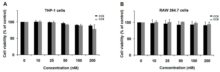
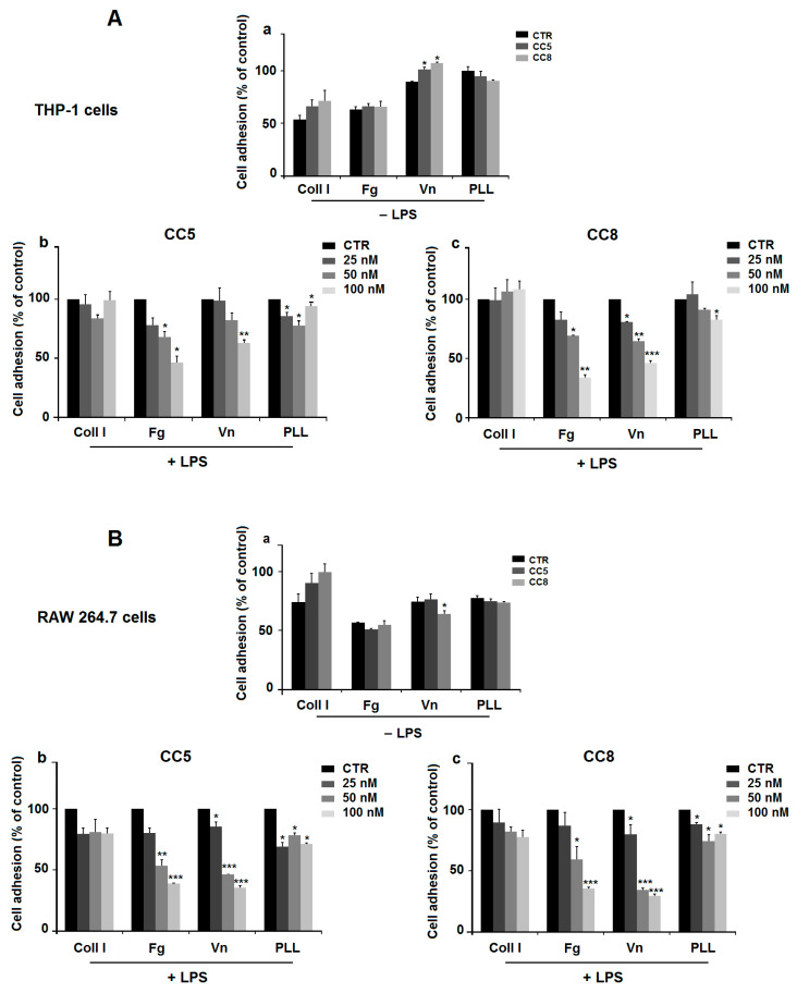
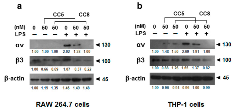
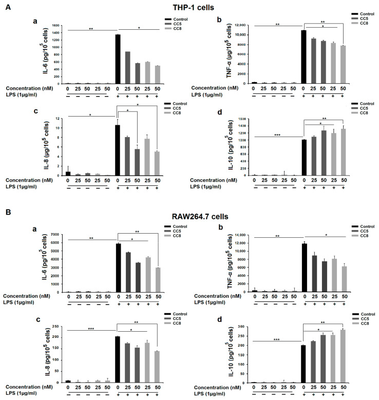
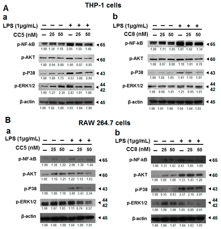
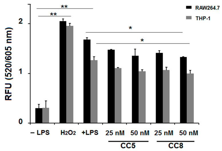
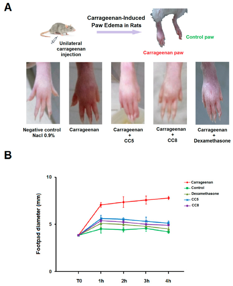
Inflammation is associated with many pathology disorders and the malignant progression of most cancers. Therefore, targeting inflammatory pathways could provide a promising strategy for disease prevention and treatment. In this study, we experimentally investigated the anti-inflammatory effect of CC5 and CC8, two disintegrin isoforms isolated from Cerastes cerastes snake venom, on LPS-stimulated macrophages, both on human THP-1 and mouse RAW264.7 cell adherence and their underlying mechanisms by measuring cytokine release levels and Western blot assay. Equally, both molecules were evaluated on a carrageenan-induced edema rat model. Our findings suggest that CC5 and CC8 were able to reduce adhesion of LPS-stimulated macrophages both on human THP-1 and mouse RAW264.7 cells to fibrinogen and vitronectin through the interaction with the αvβ3 integrin receptor. Moreover, CC5 and CC8 reduced the levels of reactive oxygen species (ROS) mediated by the NF-κB, MAPK and AKT signaling pathways that lead to decreased production of the pro-inflammatory cytokines TNF-α, IL-6 and IL-8 and increased secretion of IL-10 in LPS-stimulated THP-1 and RAW264.7 cells. Interestingly, both molecules potently exhibited an anti-inflammatory effect in vivo by reducing paw swelling in rats. In light of these results, we can propose the CC5 and CC8 disintegrins as interesting tools to design potential candidates against inflammatory-related diseases.

Keywords: cytokines; disintegrin; inflammation; signaling pathways; snake venom.


Conflict of interest statement

The authors declare no conflict of interest.

Figures

Figure 1
Figure 1
Effect of CC5 and CC8 on cell viability. (A) Effect on Human THP-1-derived macrophage viability. (B) Effect on RAW264.7 cell viability. Cells were seeded (5 × 10³/well) and cultured in presence of CC5 (black bars) or CC8 (grey bars) at different concentrations (10–200 nM) of CC5 and CC8, for 24 h. Non-treated cells were used as negative control. Cell viability was evaluated using MTT assay. Data are representative of three independent experiments performed in triplicate, expressed as mean (±SD). p < 0.05 was considered statistically significant and is indicated with asterisks over the value (* p < 0.05).
Figure 2
Figure 2
CC5 and CC8 reduce cell adhesion using cell adhesion assay. (A) Effect on THP-1-derived macrophage. (a) Cells were incubated without (black bars), or with 100 nM CC5 (dark grey bars), or with 100 nM CC8 (light grey bars) for 30 min. Cells were then allowed to attach on fibrinogen (Fg), Collagen I (Coll I), vitronectin (Vn), or poly-L-lysine (PLL). (b) Cells were stimulated with LPS (1 µg/mL) and seeded in wells coated with different extracellular matrices and incubated without or with various concentrations of CC5. (c) LPS-stimulated THP-1 cells were seeded in wells pretreated with different extracellular matrices and incubated without or with various concentrations of CC8. Non-treated cells were used as negative control. (B) Effect on RAW264.7 cells. (a) Cells were incubated without (black bars), or with 100 nM CC5 (dark grey bars), or with 100 nM CC8 (light grey bars) for 30 min. Cells were then allowed to attach on Fg, Coll I, Vn, or PLL. (b) Cells were stimulated with LPS (1 µg/mL), seeded in wells coated with different extracellular matrices and incubated without or with CC5 at various concentrations. (c) LPS-stimulated RAW264.7 cells were seeded in wells pretreated with different extracellular matrices and incubated without or with various concentrations of CC8. Non-treated cells were used as negative control. Data are representative of three independent experiments performed in triplicate, expressed as mean (±SD). p < 0.05 was considered statistically significant and is indicated with asterisks over the value (* p < 0.05, ** p < 0.01, *** p < 0.001).
Figure 3
Figure 3
CC5 and CC8 binding with αvβ3 integrin receptor on LPS-stimulated macrophages. (a) Human THP-1-derived macrophages were treated or not with LPS (1 μg/mL) in the absence and the presence of CC5 and CC8 (50 nM). (b) RAW264.7 cells were treated or not with LPS (1 μg/mL) in the absence and the presence of CC5 and CC8 (50 nM). Cell lysates were analyzed by Western blotting assay, and quantification was performed by using the software program Image J (IJ152). β-actin was used as a loading control. In the presence of LPS, integrin expression in treated cells by protein was compared to untreated cells. All data were statistically significant (p < 0.05).
Figure 4
Figure 4
Effect of CC5 and CC8 on inflammatory cytokine release in LPS-stimulated THP-1 and RAW264.7 cells using enzyme-linked immunosorbent assay. (A) Human THP-1-derived macrophages were incubated for 24 h in the absence or presence of LPS (1 µg/mL). After treatment with different concentrations of CC5 (dark grey bars) or CC8 (light grey bar), secretions of IL-6 (a), TNF-α (b), IL-8 (c), and IL-10 (d) were measured. (B) RAW264.7 cells were incubated for 24 h in the absence or presence of LPS (1 µg/mL). After treatment with different concentrations of CC5 (dark grey bars) or CC8 (light grey bars), secretions of IL-6 (a), TNF-α (b), IL-8 (c), and IL-10 (d) were measured. The negative controls (black bars) correspond to non-treated cells. Results are reported as the means ± SE of three independent experiments performed in triplicate. p < 0.05 was considered statistically significant and is indicated with asterisks over the value (* p < 0.05, ** p < 0.01, *** p < 0.001).
Figure 5
Figure 5
CC5 and CC8 modulate LPS-induced activation of NF-kB, ERK1/2, p38 MAPK, and Akt signaling pathways in LPS-stimulated THP-1 and RAW264.7 cells. (A) Cell lysates from human THP-1-derived macrophages untreated or treated with LPS (1 μg/mL) were analyzed by Western blot assay and performed to detect phosphorylated NF-kB, ERK1/2, p38 MAPK, and Akt in the presence or absence of different concentrations of CC5 (a) and CC8 (b). (B) Cell lysates from RAW264.7 untreated or treated with LPS (1 μg/mL) were analyzed by Western blot assay and performed to detect phosphorylated NF-κB, ERK1/2, p38 MAPK, and Akt in the presence or absence of different concentrations of CC5 (a) and CC8 (b). Protein band quantification was performed using the software program Image J. β-actin was used as a loading control. In LPS-stimulated cells, all data were statistically significant (p < 0.05) compared to control (untreated cells with proteins), except p-AKT in THP-1 cells with CC8 (at 25 nM), p-AKT, p-P38 in RAW264.7 cells with CC5 (at 25 nM) and p-AKT, pERK1/2 in RAW264.7 cells with CC8 (at 25 nM).
Figure 6
Figure 6
Intracellular ROS production decrease by CC5 and CC8 on LPS-stimulated THP-1 and RAW264.7 cells by measurement with intracellular ROS generation assay. RAW264.7 cells (black bars) and THP-1 cells (grey bars) were stimulated or not with LPS (1 µg/mL) and treated with different concentrations of CC5 and CC8. Untreated cells were used as negative control. H2O2 (1 mM) was used as positive control. Data are the mean of two independent experiments. p < 0.05 was considered statistically significant and is indicated with asterisks over the value (* p < 0.05, ** p < 0.01).
Figure 7
Figure 7
In vivo testing of anti-inflammatory effect of CC5 and CC8 in rats using carrageenan-induced paw edema model. Rats were randomly allocated to five groups of three rats each: Paw edema was induced by injection of carrageenan (15 mg/kg) into rat pads. CC5, CC8 (5 µg), or dexamethasone (1 mg/kg) were administrated by intraperitoneal route. The negative control group received 2.5 mL/kg of physiological solution 0.9% NaCl. (A) Macroscopic view of paw edema in different groups for 4 h following carrageenan injection. (B) The size of the edema was followed by measuring changes in paw thickness after carrageenan injection using a sliding caliper at various times (0, 1, 2, 3, and 4 h). Data are presented as means ± SD. All data were statistically significant (p < 0.05) when compared to the carrageenan group.

Similar articles

Cited by

References

    1. Germolec D.R., Shipkowski K.A., Frawley R.P., Evans E. Markers of Inflammation. Methods Mol. Biol. 2018;1803:57–79. doi: 10.1007/978-1-4939-8549-4_5. - DOI - PubMed
    1. Medzhitov R. Inflammation 2010: New adventures of an old flame. Cell. 2010;140:771–776. doi: 10.1016/j.cell.2010.03.006. - DOI - PubMed
    1. Abdulkhaleq L.A., Assi M.A., Abdullah R., Zamri-Saad M., Taufiq-Yap Y.H., Hezmee M.N.M. The crucial roles of inflammatory mediators in inflammation: A review. Vet. World. 2018;11:627–635. doi: 10.14202/vetworld.2018.627-635. - DOI - PMC - PubMed
    1. Lawrence T. The nuclear factor NF-kappaB pathway in inflammation. Cold Spring Harb. Perspect. Biol. 2019;1:a001651. doi: 10.1101/cshperspect.a001651. - DOI - PMC - PubMed
    1. Kawabata K., Tung N.H., Shoyama Y., Sugie S., Mori T., Tanaka T. Dietary Crocin Inhibits Colitis and Colitis-Associated Colorectal Carcinogenesis in Male ICR Mice. Evid. Based Complement. Altern. Med. 2012;2012:820415. doi: 10.1155/2012/820415. - DOI - PMC - PubMed

LinkOut - more resources