. 2023 Aug 5;24(15):12455.
doi: 10.3390/ijms241512455.

TET Family Members Are Integral to Porcine Oocyte Maturation and Parthenogenetic Pre-Implantation Embryogenesis

Affiliations

TET Family Members Are Integral to Porcine Oocyte Maturation and Parthenogenetic Pre-Implantation Embryogenesis

Fan Chen et al. Int J Mol Sci. .

Abstract

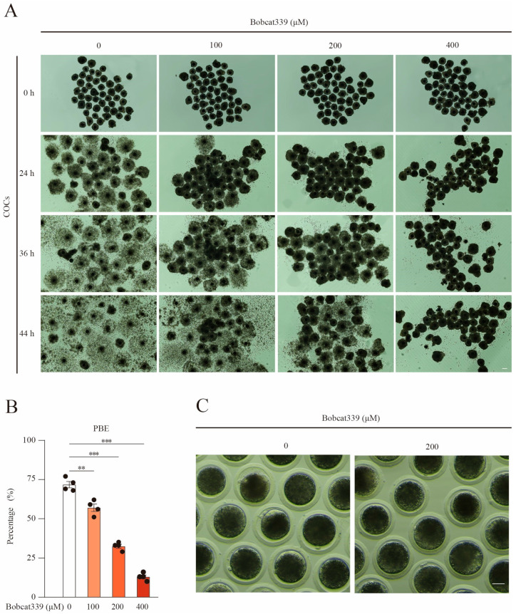
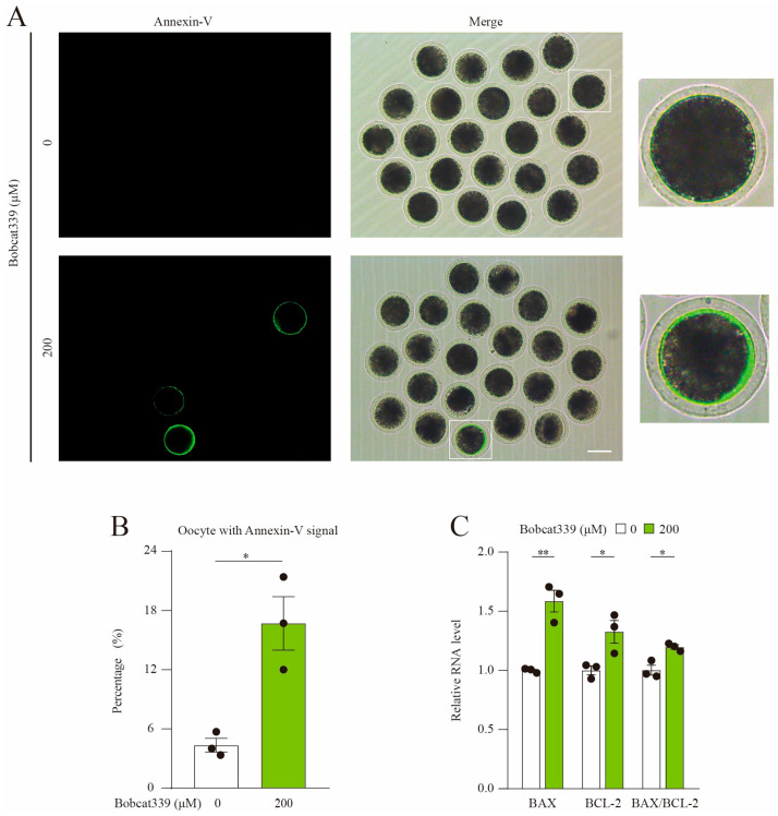
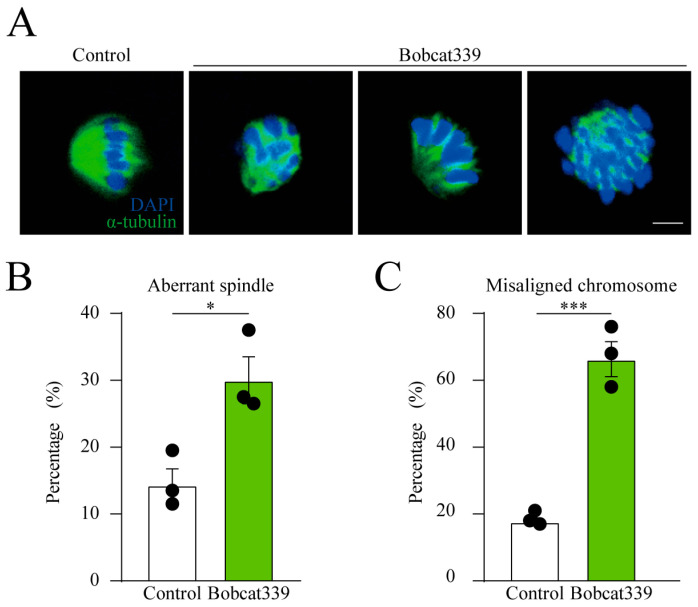
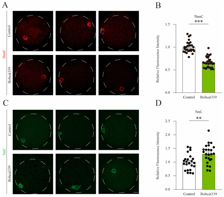
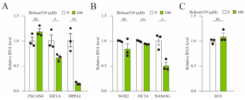
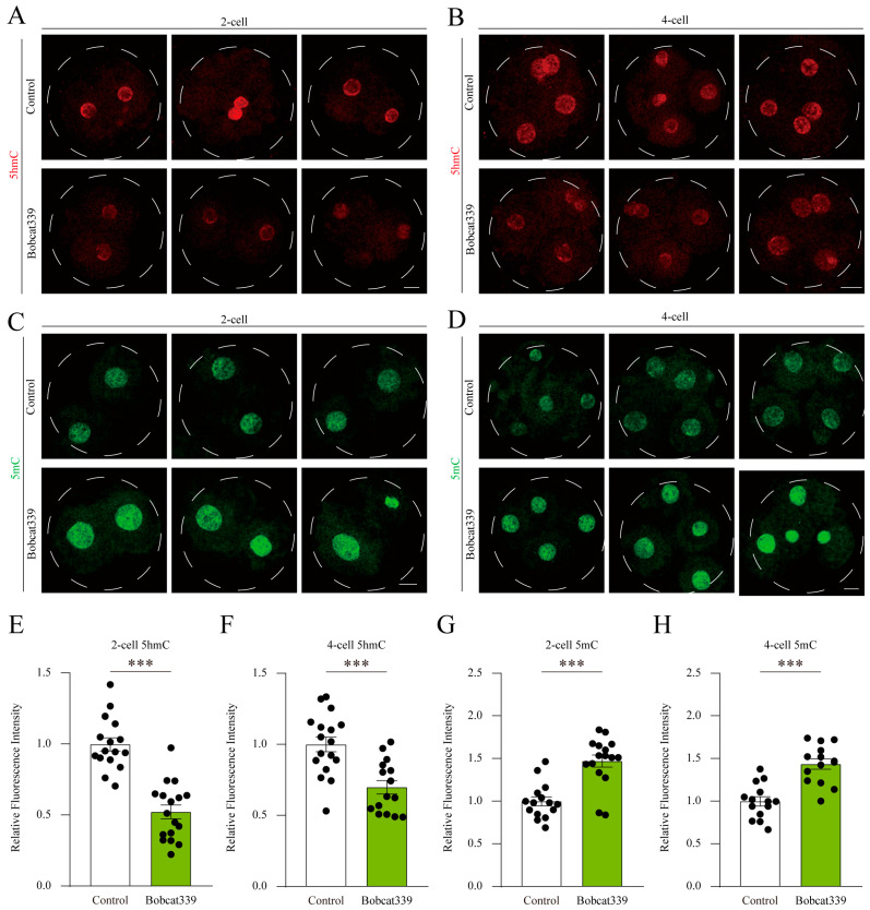
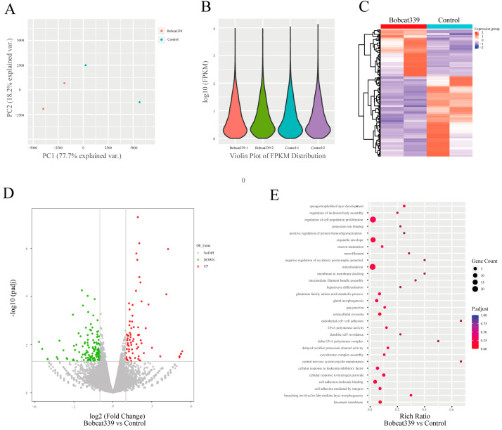
The ten-eleven translocation (TET) enzyme family, which includes TET1/2/3, participates in active DNA demethylation in the eukaryotic genome; moreover, TET1/2/3 are functionally redundant in mice embryos. However, the combined effect of TET1/2/3 triple-gene knockdown or knockout on the porcine oocytes or embryos is still unclear. In this study, using Bobcat339, a specific small-molecule inhibitor of the TET family, we explored the effects of TET enzymes on oocyte maturation and early embryogenesis in pigs. Our results revealed that Bobcat339 treatment blocked porcine oocyte maturation and triggered early apoptosis. Furthermore, in the Bobcat339-treated oocytes, spindle architecture and chromosome alignment were disrupted, probably due to the huge loss of 5-hydroxymethylcytosine (5hmC)and concurrent increase in 5-methylcytosine (5mC). After Bobcat339 treatment, early parthenogenetic embryos exhibited abnormal 5mC and 5hmC levels, which resulted in compromised cleavage and blastocyst rate. The mRNA levels of EIF1A and DPPA2 (ZGA marker genes) were significantly decreased, which may explain why the embryos were arrested at the 4-cell stage after Bobcat339 treatment. In addition, the mRNA levels of pluripotency-related genes OCT4 and NANOG were declined after Bobcat339 treatment. RNA sequencing analysis revealed differentially expressed genes in Bobcat339-treated embryos at the 4-cell stage, which were significantly enriched in cell proliferation, cell component related to mitochondrion, and cell adhesion molecule binding. Our results indicated that TET proteins are essential for porcine oocyte maturation and early embryogenesis, and they act by mediating 5mC/5hmC levels and gene transcription.

Keywords: 5-hydroxymethylcytosine; 5-methylcytosine; Bobcat339; TET proteins; embryo; oocyte; pig.


Conflict of interest statement

The authors declare no conflict of interest.

Figures

Figure 1
Figure 1
Bobcat339 treatment impaired porcine oocyte maturation. (A) Representative images of cumulus oocyte complexes (COCs) in control and Bobcat339-treated (0, 100, 200, and 400 μM) oocytes after culturing in vitro for 24, 36, and 44 h. Good expansion of cumulus cells was observed in control COCs, whereas they exhibited less expansion and were more adhesive in the Bobcat339-treated group. Scale Bar = 50 μm. (B) The percentage of the PBE was significantly decreased after treatment with 100 (** p < 0.01), 200 (*** p < 0.001), and 400 μM (*** p < 0.001) Bobcat339. The results are shown as the mean ± SEM from at least three independent experiments. (C) Representative images of PBE in control and 200 μM Bobcat339-treated oocytes after culturing in vitro for 44 h. Scale Bar = 50 μm.
Figure 2
Figure 2
Bobcat339 treatment induced the early apoptosis of porcine oocytes. (A) Early apoptotic fluorescent signals in control and Bobcat339-treated oocytes. Annexin-V, green; Scale Bar = 100 μm. (B) The proportion of the oocytes with Annexin-V signal was calculated in control and Bobcat339-treated oocytes. (C) qRT-PCR revealed the mRNA levels of BAX, BCL-2 and BAX/BCL-2 in control and Bobcat339-treated oocytes. * p < 0.05; ** p < 0.01. The results are shown as the mean ± SEM from at least three independent experiments.
Figure 3
Figure 3
Influence of Bobcat339 on spindle morphology and chromosome alignment. (A) Images delineating spindle morphology and chromosome alignment in control and Bobcat339-treated oocytes (200 μM Bobcat339). Blue, chromosomes; Green, α-tubulin; Scale Bar = 20 μm. (B) The percentage of aberrant spindle was significantly increased in Bobcat339-treated oocytes. (C) The proportion of misaligned chromosome was significantly elevated in Bobcat339-treated oocytes. * p < 0.05; *** p < 0.001. The results are shown as the mean ± SEM from at least three independent experiments.
Figure 4
Figure 4
Bobcat339 treatment altered 5mC and 5hmC levels in porcine oocytes. The COCs in control and Bobcat339-treated groups were cultured in the IVM medium supplemented with 1 mM IBMX, to maintain the meiotic GV arrest. (A) The immunofluorescent staining of 5hmC in porcine oocytes. Red, 5hmC; Scale Bar = 20 μm. (B) The fluorescence intensity of 5hmC in control and Bobcat339-treated oocytes. (C) Immunofluorescent staining of 5mC in control and Bobcat339-treated oocytes. Green, 5mC; Scale Bar = 20 μm. (D) Average fluorescence intensity of 5mC in control and Bobcat339-treated oocytes. ** p < 0.01; *** p < 0.001. The results are shown as the mean ± SEM from at least three independent experiments.
Figure 5
Figure 5
Bobcat339 treatment hampered early embryo development in pigs. (A) Images delineating blastocyst formation on day 6 of parthenogenetically activated porcine embryos in control and Bobcat339-treated (25, 50, and 100 μM) groups. Scale Bar = 100 μm. (B) The proportions of activated oocytes that developed to the blastocyst stage were calculated between control and Bobcat339-treated groups. (C) The proportions of embryos at various stages were calculated on day 3 in control and Bobcat339-treated (25, 50, and 100 μM Bobcat339) groups. ns (no significance) represents p > 0.05; ** p < 0.01; *** p < 0.001. The results are shown as the mean ± SEM from at least three independent experiments.
Figure 6
Figure 6
Bobcat339 treatment blocked the expression of ZGA and pluripotency genes. (A) The mRNA levels of EIF1A and DPPA2 were significantly decreased in Bobcat339-treated embryos. (B) The mRNA levels of OCT4 and NANOG were reduced in Bobcat339-treated embryos; the mRNA levels of SOX2 exhibited no change. (C) The mRNA levels of H19 were not affected after Bobcat339 treatment. ns (no significance) represents p > 0.05; * p < 0.05; ** p < 0.01. The results are shown as the mean ± SEM from at least three independent experiments.
Figure 7
Figure 7
Bobcat339 treatment affected 5mC and 5hmC levels in porcine embryos. (A,B) The immunofluorescence staining of 5hmC in embryos at the 2- and 4-cell stages in control and Bobcat339-treated groups. Red, 5hmC; Scale Bar = 20 μm. (C,D) The immunofluorescence staining of 5mC in embryos at the 2- and 4-cell stages in control and Bobcat339-treated groups. Green, 5mC; Scale Bar = 20 μm. (E,F) The fluorescence intensity of 5hmC was calculated in embryos at the 2- and 4-cell stages in control and Bobcat339-treated groups. (G,H) Average fluorescence intensity of 5mC was recorded in embryos in control and Bobcat339-treated groups at the 2- and 4-cell stages. The results are shown as the mean ± SEM from at least three independent experiments. *** p < 0.001.
Figure 8
Figure 8
The different gene expression patterns between control and Bobcat339-treated embryos. (A) Principal component analysis of mRNA level between control and Bobcat339-treated embryos at the 4-cell stage. (B) Violin plot of gene expression and distribution between control and Bobcat339-treated groups. (C) Hierarchical cluster of differentially expressed genes (DEGs; p < 0.05 and |log2 fold change| > 1) between control and Bobcat339-treated embryos at the 4-cell stage. The color scale of the heatmap represents the expression levels. (D) Volcano plot of DEGs between control and Bobcat339-treated embryos at the 4-cell stage. Red and green dots represent up- and down regulated genes, respectively. (E) Gene Ontology analysis revealed that the DEGs were enriched in certain biological processes, molecular functions, and cellular components.

Similar articles

Cited by

References

    1. Kennedy A.J., Sweatt J.D. Drugging the methylome: DNA methylation and memory. Crit. Rev. Biochem. Mol. Biol. 2016;51:185–194. doi: 10.3109/10409238.2016.1150958. - DOI - PMC - PubMed
    1. Ito S., Shen L., Dai Q., Wu S.C., Collins L.B., Swenberg J.A., He C., Zhang Y. Tet proteins can convert 5-methylcytosine to 5-formylcytosine and 5-carboxylcytosine. Science. 2011;333:1300–1303. doi: 10.1126/science.1210597. - DOI - PMC - PubMed
    1. He Y.F., Li B.Z., Li Z., Liu P., Wang Y., Tang Q., Ding J., Jia Y., Chen Z., Li L., et al. Tet-mediated formation of 5-carboxylcytosine and its excision by TDG in mammalian DNA. Science. 2011;333:1303–1307. doi: 10.1126/science.1210944. - DOI - PMC - PubMed
    1. Wu X., Zhang Y. TET-mediated active DNA demethylation: Mechanism, function and beyond. Nat. Rev. Genet. 2017;18:517–534. doi: 10.1038/nrg.2017.33. - DOI - PubMed
    1. Shi D.Q., Ali I., Tang J., Yang W.C. New Insights into 5hmC DNA Modification: Generation, Distribution and Function. Front. Genet. 2017;8:100. doi: 10.3389/fgene.2017.00100. - DOI - PMC - PubMed