. 2023 Aug 7;24(15):12517.
doi: 10.3390/ijms241512517.

Robust ParB Binding to Half- parS Sites in Pseudomonas aeruginosa-A Mechanism for Retaining ParB on the Nucleoid?

Affiliations

Robust ParB Binding to Half- parS Sites in Pseudomonas aeruginosa-A Mechanism for Retaining ParB on the Nucleoid?

Adam Kawalek et al. Int J Mol Sci. .

Abstract

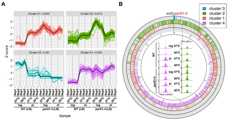
Chromosome segregation in Pseudomonas aeruginosa is assisted by the tripartite ParAB-parS system, composed of an ATPase (ParA), a DNA-binding protein (ParB) and its target parS sequence(s). ParB forms a nucleoprotein complex around four parSs (parS1-parS4) that overlaps oriC and facilitates relocation of newly synthesized ori domains inside the cells by ParA. Remarkably, ParB of P. aeruginosa also binds to numerous heptanucleotides (half-parSs) scattered in the genome. Here, using chromatin immunoprecipitation-sequencing (ChIP-seq), we analyzed patterns of ParB genome occupancy in cells growing under conditions of coupling or uncoupling between replication and cell division processes. Interestingly, a dissipation of ParB-parS complexes and a shift of ParB to half-parSs were observed during the transition from the exponential to stationary phase of growth on rich medium, suggesting the role of half-parSs in retaining ParB on the nucleoid within non-dividing P. aeruginosa cells. The ChIP-seq analysis of strains expressing ParB variants unable to dislocate from parSs showed that the ParB spreading ability is not required for ParB binding to half-parSs. Finally, a P. aeruginosa strain with mutated 25 half-parSs of the highest affinity towards ParB was constructed and analyzed. It showed altered ParB coverage of the oriC region and moderate changes in gene expression. Overall, this study characterizes a novel aspect of conserved bacterial chromosome segregation machinery.

Keywords: ParB distribution; Pseudomonas aeruginosa; chromosome segregation; half-parSs; partitioning proteins.


Conflict of interest statement

The authors declare no conflict of interest. The funders had no role in the design of the study; in the collection, analyses, or interpretation of data; in the writing of the manuscript or in the decision to publish the results.

Figures

Figure 1
Figure 1
ParB binding to the P. aeruginosa chromosome is dependent on the growth conditions. Growth of Pae PAO1161 (WT) strain in LB or M9 medium +0.5% glucose at 30 °C or 37 °C presented as (A) mean optical density at 600 nm ± SD from three cultures and (B) mean number of colony-forming units (CFUs) per mL ± SD assayed for three cultures. (C) ChIP-seq analysis of ParB binding to genome assayed using WT strain cells from cultures in LB or M9 medium, grown at 30 or 37 °C, at two time points representing exponential and stationary phases of culture growth (indicated on (A) with red lines). Track data represent normalized coverage of the genome with ChIP-seq reads (binned at 200 bp and averaged from two ChIP-seq replicates). Color of coverage track represents different growth conditions, whereas track background indicates the phase of culture growth. Track height was set at 10% of max. coverage signal of the outer track. Short bars below each track represent the distribution of detected ChIP-seq peaks for these samples, colored according to the peak fold enrichment (FE).
Figure 2
Figure 2
Formation of ParB–parS complexes in Pae PAO1161 cells depends on the growth conditions. (A,B) Coverage of the genome regions encompassing the parS1parS4 in the PAO1161 genome in the ChIP-seq samples for the indicated strains/culture conditions. The histograms show normalized coverage with reads (averaged for two biological replicates). (CF) Microscopic analysis of PAO1161 YFP–ParB producing cells grown in (C) LB medium at 37 °C or (E) M9 medium with 0.5% glucose at 37 °C at different time points of culture growth. On the merged images, the fluorescence signal was enhanced, and the cell contour was false-colored in red. Marked areas were copied and enlarged (C), and the asterisk at the 24 h panel indicates a rare cell with foci in this conditions. (D,F) Histograms showing distribution of the number of YFP–ParB foci per cell, detected in fluorescence images using MicrobeJ ver 5.13m.
Figure 3
Figure 3
Redistribution of ParB from complex around parSs to half-parS sites with changes in growth conditions. (A) Number of ParB ChIP-seq peaks for samples derived from the indicated strain and growth conditions, categorized according to their fold enrichment (FE). (B) Correlation heatmap between different ChIP-seq peak sets, based on occupancy and MACS2 scores. (C) Percentage of ChIP-seq peaks containing GTTCCAC or GTTTCAC (half-parS) motifs ± 50 bp from the summit. The analysis was performed for all peaks (upper panel) and for peaks with FE > 3 (bottom panel). (D) Percentage of ChIP-seq reads mapping to genome regions encompassing parS1–4 or half-parSs in different samples. Reads mapping to parSs ± 300 bp, ParB spreading zone without parSs and regions with half-parSs (GTTCCAC and GTTTCAC ± 300 bp, altogether 11.6% of the genome) were counted, and background (based on the amount of reads mapping to the remaining genome regions) was subtracted. Data for ChIP-seq replicates were averaged. (E) Read coverage around GTTCCAC motifs in the PAO1161 genome in ParB ChIP-seq samples. Individual lines in the heatmap represent normalized read counts for regions ±300 bp around a motif. The data were sorted in descending order of median coverage value and represent averaged value for two biological replicates. Plots indicate median coverage over the analyzed regions.
Figure 4
Figure 4
The preference of ParB binding to individual half-parS motifs is unaffected in cells grown in different media, at the tested temperatures and does not depend on the phase of growth. Coverage data for all 971 GTTCCAC motifs present in the PAO1161 genome were extracted from ChIP-seq data, normalized (Z-score) and subjected to k-means clustering. (A) ChIP-seq read coverage profiles for motifs in each cluster. The y-axis represents the difference (in standard deviations) between coverage of a particular motif in individual ChIP-seq samples and the mean coverage calculated for this motif in all samples. Thick black lines represent the cluster means. (B) Distribution of motifs from the four clusters in the PAO1161 genome. Inset represents an example of motif coverage profiles for selected motifs from cluster 2 and 4 in ChIP-seq samples derived from strains/indicated growth conditions.
Figure 5
Figure 5
Effects of mutations in the arginine rich patch of ParB and deletion of the C-terminus on ParB interactions with DNA, assessed using ChIP-seq. (A) Coverage of regions encompassing the parS1–parS4 in the PAO1161 genome in the ChIP-seq samples for the indicated strains. The histograms show normalized coverage with reads (averaged for two biological replicates). (B) Distribution of ChIP-seq peaks for the analyzed strains in the genome. Bar height and colors indicate peak fold enrichment (FE). (C) Number of peaks with and without GTTCCAC or GTTTCAC motifs ± 50 bp from the summit. The analysis was performed only for peaks with FE > 3. (D) Read coverage around GTTCCAC and GTTTCAC motifs in the PAO1161 genome in the ParB ChIP-seq samples of different strains. Individual lines in the heatmap represent normalized read counts for regions ±300 bp around a motif. The data were sorted in descending order of median coverage value and represent averaged value for two biological replicates. Plots indicate median coverage over the analyzed region for the two motifs.
Figure 6
Figure 6
ParB binding to half-parS plays an indirect role in gene expression regulation in P. aeruginosa. (A) Circular plot indicating genomic localization of peaks identified in WT and half-parS25 strains (tracks 1 and 2) and peaks identified only in one of the strains (track 3 and 4). The positions of half-parS motifs modified in half-parS25 strain are colored with violet. The outer ring indicates 67 genes identified as differentially expressed in both RNA-seq experiments, colored according to the expression change trend. Labels indicate PAO1 gene IDs, or PAO1161 IDs (D3C65_), for genes annotated only in the PAO1161 genome. (B) Comparison of ChIP-seq peaks identified in WT and half-parS25 strains. (C) Comparison of sets of differentially expressed genes identified in two independent RNA-seq analyses. (D) Effect of the half-parS mutations on the transcription of surrounding genome fragments. Reads mapping to genome fragments ± 1000 bp from GTTCCAC motifs were counted in RNA-seq sample, and normalized for the total read count, and the ratio between WT and half-parS25 samples was calculated. Dots indicate outliers. Statistical significance between the two groups was evaluated using the Wilcoxon rank sum test.

Similar articles

Cited by

References

    1. Reyes-Lamothe R., Sherratt D.J. The bacterial cell cycle, chromosome inheritance and cell growth. Nat. Rev. Microbiol. 2019;17:467–478. doi: 10.1038/s41579-019-0212-7. - DOI - PubMed
    1. Badrinarayanan A., Le T.B.K., Laub M.T. Bacterial chromosome organization and segregation. Annu. Rev. Cell Dev. Biol. 2015;31:171–199. doi: 10.1146/annurev-cellbio-100814-125211. - DOI - PMC - PubMed
    1. den Blaauwen T. Prokaryotic cell division: Flexible and diverse. Curr. Opin. Microbiol. 2013;16:738–744. doi: 10.1016/j.mib.2013.09.002. - DOI - PubMed
    1. Bouet J.-Y., Stouf M., Lebailly E., Cornet F. Mechanisms for chromosome segregation. Curr. Opin. Microbiol. 2014;22:60–65. doi: 10.1016/j.mib.2014.09.013. - DOI - PubMed
    1. Kawalek A., Wawrzyniak P., Bartosik A.A., Jagura-Burdzy G. Rules and exceptions: The role of chromosomal ParB in DNA segregation and other cellular processes. Microorganisms. 2020;8:105. doi: 10.3390/microorganisms8010105. - DOI - PMC - PubMed

MeSH terms