. 2023 Aug 7;24(15):12525.
doi: 10.3390/ijms241512525.

Molecular Mechanisms of Endothelialitis in SARS-CoV-2 Infection: Evidence for VE-Cadherin Cleavage by ACE2

Affiliations

Molecular Mechanisms of Endothelialitis in SARS-CoV-2 Infection: Evidence for VE-Cadherin Cleavage by ACE2

Laurence Bouillet et al. Int J Mol Sci. .

Abstract

Long COVID-19 syndrome appears after Severe Acute Respiratory Syndrome-Corona Virus (SARS-CoV-2) infection with acute damage to microcapillaries, microthrombi, and endothelialitis. However, the mechanisms involved in these processes remain to be elucidated. All blood vessels are lined with a monolayer of endothelial cells called vascular endothelium, which provides a the major function is to prevent coagulation. A component of endothelial cell junctions is VE-cadherin, which is responsible for maintaining the integrity of the vessels through homophilic interactions of its Ca++-dependent adhesive extracellular domain. Here we provide the first evidence that VE-cadherin is a target in vitro for ACE2 cleavage because its extracellular domain (hrVE-ED) contains two amino acid sequences for ACE2 substrate recognition at the positions 256P-F257 and 321PMKP-325L. Indeed, incubation of hrVE-ED with the active ectopeptidase hrACE2 for 16 hrs in the presence of 10 μM ZnCl2 showed a dose-dependent (from 0.2 ng/μL to 2 ng/μL) decrease of the VE-cadherin immunoreactive band. In vivo, in the blood from patients having severe COVID-19 we detected a circulating form of ACE2 with an apparent molecular mass of 70 kDa, which was barely detectable in patients with mild COVID-19. Of importance, in the patients with severe COVID-19 disease, the presence of three soluble fragments of VE-cadherin (70, 62, 54 kDa) were detected using the antiEC1 antibody while only the 54 kDa fragment was present in patients with mild disease. Altogether, these data clearly support a role for ACE2 to cleave VE-cadherin, which leads to potential biomarkers of SARS-CoV-2 infection related with the vascular disease in "Long COVID-19".

Keywords: ACE2; SARS-CoV-2 infection; VE-cadherin; endothelium; long COVID syndrome.


Conflict of interest statement

The authors declare no conflict of interest.

Figures

Figure 1
Figure 1
The EJ are composed of a transmembrane protein (VE-cadherin). (a) Schematic representation of a capillary composed of three ECs. The capillary is surrounded by pericytes. The overlap of two ECs is the EJ. (b) Electron microscopy of the endothelial junction. The dashed white line delineates two ECs. (c) Extracellular domain of VE-cadherin showing the hydrophobic amino acid W49 and W51 responsible for cohesion of ECs. (d) Coomassie-stained SDS-PAGE showing that human recombinant extracellular domain of VE-cadherin with an apparent mass of 90 kDa and immunoblotted with VE-cadherin antibody (clone BV9) not treated (left lane) or treated (right lane) with PGNase-F. (e) Schematic representation of VE-cadherin extracellular domain with the epitopes of the antibodies (Anti-EC1 and the clone BV9) against human VE-cadherin. (f) Immunoblotting of human recombinant VE-cadherin with the antibodies clone BV9 and anti-EC1. The positions of molecular mass standards (in kDa) are shown at the left. These experiments were repeated at least four times in a similar configuration.
Figure 2
Figure 2
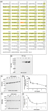
The extracellular domain of VE-cadherin is a substrate for the ectopeptidase ACE2: (a) VE-cadherin sequence. The yellow color highlights the amino acid sequence of the extracellular domain of the protein. The putative consensus site for the ACE2 cleavage is highlighted in red. (b) Representative western blot showing the human recombinant ACE2 (hrACE2) analyzed with the antibody directed against ACE2 extracellular domain. The positions of molecular mass standards (in kDa) are shown at the left (c,d). Representative western blots of VE-cadherin extracellular domain incubated overnight at room temperature with increasing concentrations of hrACE2 (0.1 to 4 ng/mL) in 50 mM 2-(N-Morpholino)-ethane sulfonic acid (MES), 300 mM NaCl, 10 μM ZnCL2, 0.01% Brig 35, pH 6.5). The samples were analyzed by SDS-PAGE and immunoblotting. The immunoreactive band was detected with the monoclonal anti VE-cadherin antibody (BV9) or the rabbit polyclonal anti-EC1 antibody (anti EC1). (e,f) Densitometric analysis of the 90 kDa band using ImageJ software showed a dose-dependent decrease in VE-cadherin band intensity. This experiment was performed four times (n = 4) under similar conditions with comparable results.
Figure 3
Figure 3
Analysis of circulating ACE2 in blood from patients with SARS-CoV-2 infection. (a) Blood samples from COVID-19 patients were analyzed by SDS-PAGE and immunoblotting. Blood samples were diluted serially from 1 to 10 followed by a dilution of 1 to 2.5. 15 μL of sample preparations were loaded onto an SDS-PAGE and then transferred onto a nitrocellulose membrane. Proteins on the blots were visualized by Ponceau staining. The positions of molecular mass standards (in kDa) are shown at the left. The membranes were incubated with the anti-ACE2 antibody (5 μg/mL) followed by incubation with the anti-mouse peroxidase antibody. The arrow on the left-hand side shows the immunoreactive band corresponding to cACE2. (b) Densitometry analysis of the immunoreactive band corresponding to cACE2 to compare the groups (n = 4 for mild infection and n = 5 for severe infection). Error bars represent mean ± SE of means. p values from analysis of variance were assessed using the Mann-Whitney test (* p < 0.05). These experiments were repeated at least four times in a similar configuration.
Figure 4
Figure 4
Analysis of the soluble forms of VE-cadherin in COVID-19 patient blood. (a) Blood samples from COVID-19 patients were analyzed by SDS-PAGE and immunoblotting. Proteins on the blots were visualized by Ponceau staining. Blood samples were diluted serially from 1 to 10 followed by a dilution of 1 to 5 (total dilution 1:50). 15 μL of sample preparations were loaded onto the gel and then immunoblotted with the anti-VE-cadherin antibody (EC1 1:250) overnight at 4 °C followed by an incubation with the goat anti-rabbit peroxidase antibody (1:2000). Arrows indicate the apparent molecular masses of the immunoreactive VE-cadherin. The positions of molecular mass standards (in kDa) are shown at the left. This experiment is representative of three independent experiments. (bd). Densitometry analysis to compare the groups (n = 4 for mild infection and n = 5 for severe infection) for the three different forms of soluble VE-cadherin 70 kDa (b), 62 kDa (c), 54 kDa (d). The differences between patient groups were assessed as being significant using Mann-Whitney analysis (* p < 0.05). These experiments were repeated at least four times in a similar configuration.
Figure 5
Figure 5
Proposed model of Pulmonary Endothelial dysfunction in COVID patients: In the pulmonary environment, the ECs are surrounded by pneumocytes (P1, and P2) and macrophages that are responsible for the cytokine storm. ACE2 is present on ECs as a transmembrane protein whose catalytic site is outside the cells (ectopeptidase). ACE2 can be cleaved by ADAM 17 and leads to the generation of a circulating active form of ACE2. VE-cadherin is a transmembrane protein exclusively expressed in ECs, which can be subjected to post-translational modifications including tyrosine phosphorylation upon cytokine challenge. This covalent modification will lead to a conformational change in the protein structure, which presents a high sensitivity to proteases. Thus, the ACE2 enzyme will act directly to its potential site of cleavage generating the fragments of VE-cadherin seen in the patient’s blood.

References

    1. Zanini G., Selleri V., Roncati L., Coppi F., Nasi M., Farinetti A., Manenti A., Pinti M., Mattioli A.V. Vascular “Long COVID”: A new vessel disease? Angiology. 2023:33197231153204. doi: 10.1177/00033197231153204. - DOI - PMC - PubMed
    1. Wu C., Chen X., Cai Y., Xia J., Zhou X., Xu S., Huang H., Zhang L., Zhou X., Du C., et al. Risk factors associated with acute respiratory distress syndrome and death in patients with Coronavirus disease 2019 pneumonia in Wuhan, China. JAMA Intern. Med. 2020;180:934–943. doi: 10.1001/jamainternmed.2020.0994. - DOI - PMC - PubMed
    1. WHO Coronavirus (COVID-19) Dashboard. [(accessed on 1 January 2020)]. Available online: https://covid19.who.int.
    1. Varga Z., Flammer A.J., Steiger P., Haberecker M., Andermatt R., Zinkernagel A.S., Mehra M.R., Schuepbach R.A., Ruschitzka F., Moch H. Endothelial cell infection and endotheliitis in COVID-19. Lancet. 2020;395:1417–1418. doi: 10.1016/S0140-6736(20)30937-5. - DOI - PMC - PubMed
    1. Ackermann M., Verleden S.E., Kuehnel M., Haverich A., Welte T., Laenger F., Vanstapel A., Werlein C., Stark H., Tzankov A., et al. Pulmonary vascular endothelialitis, thrombosis, and angiogenesis in COVID-19. N. Engl. J. Med. 2020;383:120–128. doi: 10.1056/NEJMoa2015432. - DOI - PMC - PubMed

MeSH terms