Review
. 2023 Jul 27;28(15):5686.
doi: 10.3390/molecules28155686.

Significance of Five-Membered Heterocycles in Human Histone Deacetylase Inhibitors

Affiliations
Review

Significance of Five-Membered Heterocycles in Human Histone Deacetylase Inhibitors

Anton Frühauf et al. Molecules. .

Abstract

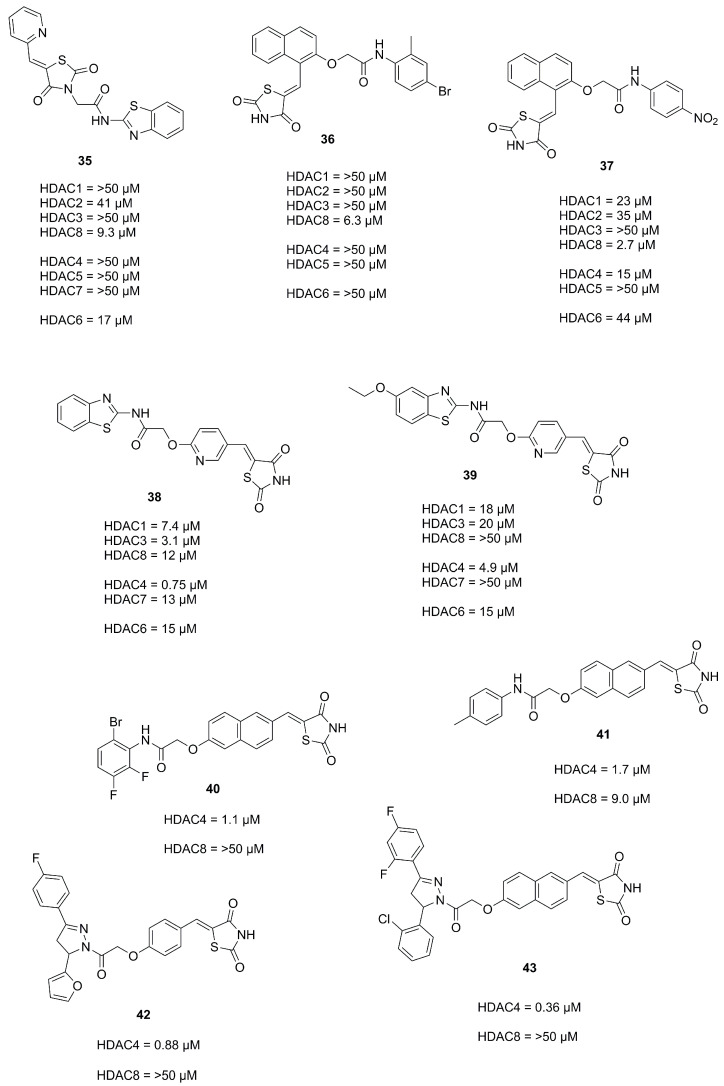
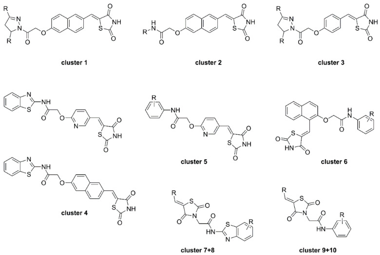
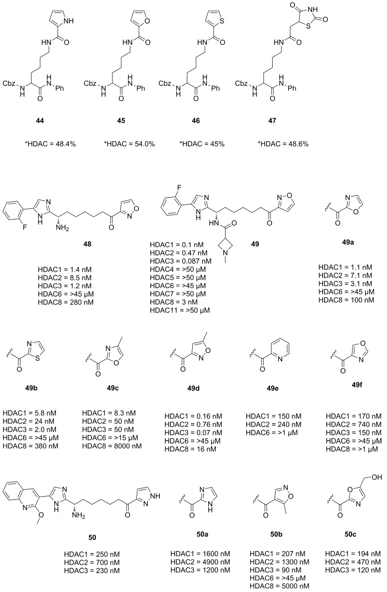
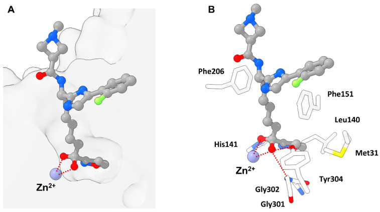
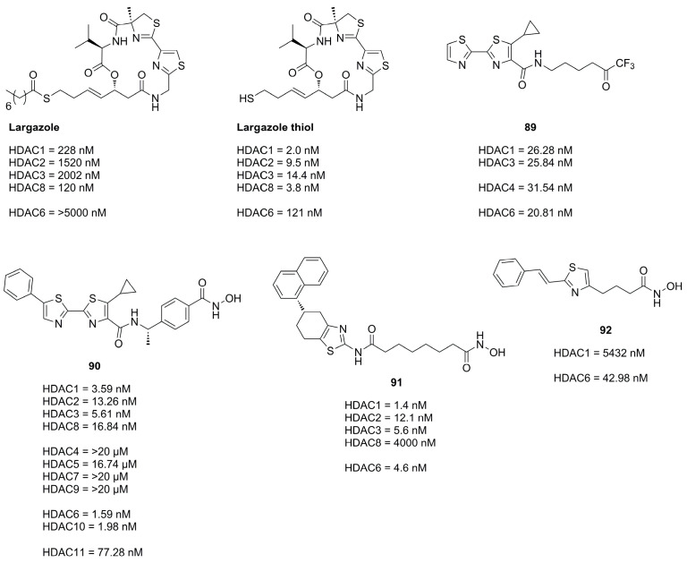
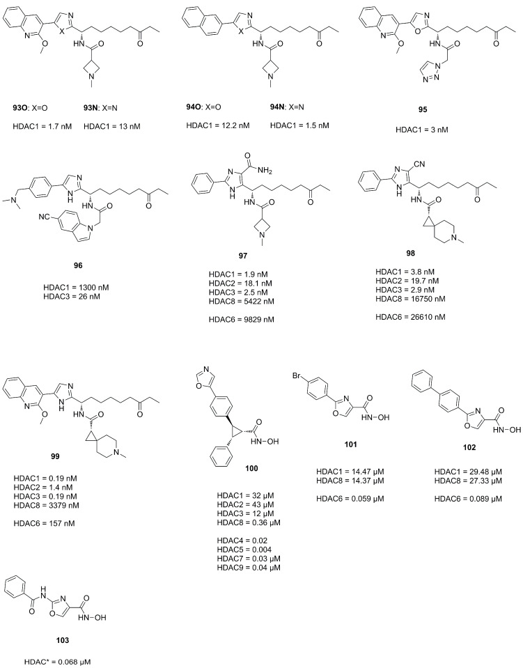
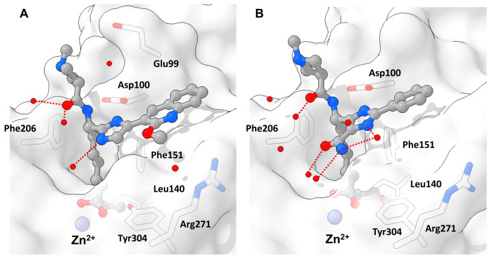
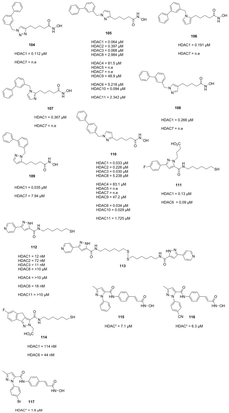
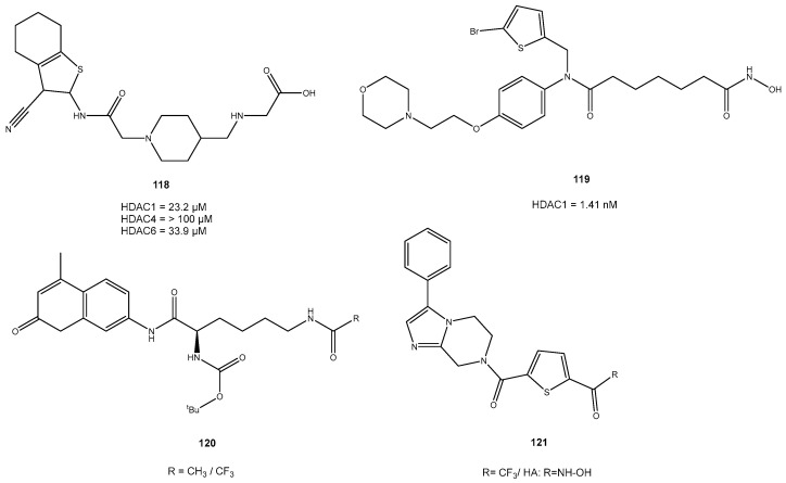
Five-membered heteroaromatic rings, in particular, have gained prominence in medicinal chemistry as they offer enhanced metabolic stability, solubility and bioavailability, crucial factors in developing effective drugs. The unique physicochemical properties and biological effects of five-membered heterocycles have positioned them as key structural motifs in numerous clinically effective drugs. Hence, the exploration of five-ring heterocycles remains an important research area in medicinal chemistry, with the aim of discovering new therapeutic agents for various diseases. This review addresses the incorporation of heteroatoms such as nitrogen, oxygen and sulfur into the aromatic ring of these heterocyclic compounds, enhancing their polarity and facilitating both aromatic stacking interactions and the formation of hydrogen bonds. Histone deacetylases are present in numerous multiprotein complexes within the epigenetic machinery and play a central role in various cellular processes. They have emerged as important targets for cancer, neurodegenerative diseases and other therapeutic indications. In histone deacetylase inhibitors (HDACi's), five-ring heterocycles perform various functions as a zinc-binding group, a linker or head group, contributing to binding activity and selective recognition. This review focuses on providing an up-to-date overview of the different five-membered heterocycles utilized in HDACi motifs, highlighting their biological properties. It summarizes relevant publications from the past decade, offering insights into the recent advancements in this field of research.

Keywords: active pharmaceutical ingredient; active substance optimization; drug design; drug development; histone deacetylase inhibitors.


Conflict of interest statement

The authors declare no conflict of interest.

Figures

Figure 1
Figure 1
Schematic classification of HDAC isozymes into class I, IIa, IIb and IV. Indicated are catalytic domain size (blue box), protein length (size) and localization.
Figure 2
Figure 2
Approved HDAC inhibitors with indicated structural features. Cap group, linker and ZBG are labelled in green, blue and black, respectively.
Figure 3
Figure 3
Exemplary binding pose of a benzamide HDACi with indicated structural features inside a HDAC2 binding pocket (PDB-ID: 4LY1, 1.57 Å) [15] illustrated using the UCSF ChimeraX visualization software (version ChimeraX-1.4) [16,17]. The inhibitor and the amino acid side chains are displayed in the ball and stick or the stick style and are colored by element or heteroatom, respectively. (A) Clipped binding pocket indicating the canonical binding pocket occupied by the linker and ZBG motive and the acetate release channel occupied by the foot pocket motive. The zinc ion is shown as bluish sphere and interacts with the ZBG group via indicated dotted red lines. (B) Most prominent residues and interactions in a typical class I binding site. The greatest energy contribution is provided by the interaction between the ZBG group and the zinc ion as well as hydrogen bond stabilization between Tyr304 and the carbonyl functionality of the HDACi, indicated as dotted red lines. The substrate tunnel is composed of hydrophobic residues of which Phe151 and Phe206 can be exploited for π-stacking interactions.
Figure 4
Figure 4
Schematic illustration of bond lengths (Å), bond angles (°), dipole moments (D) and calculated hydrogen bond acceptor strengths in kcal/mol indicated in blue.
Figure 5
Figure 5
Properties and reactivities of 1,2,4-oxadiazole derivatives. The electron-withdrawing nature of azoximes is indicated by diol formation of 3. C3 and C5 reactivity is illustrated with 4 and 5 in the presence of electron withdrawing groups and can be rationalized with mesomeric structures of 6 and 7.
Figure 6
Figure 6
Representation of TMP269 bound to HDAC7 (PDB-ID: 3ZNR, 2.35 Å) illustrated using the UCSF ChimeraX visualization software (version ChimeraX-1.4) [16,17]. The inhibitor and the amino acid side chains are displayed in the ball and stick or the stick style and are colored by element or heteroatom, respectively. (A) Notable is the larger binding site of class IIa HDACs compared to other HDAC classes. Possible interactions between TMP269 and the zinc ion are indicated as dotted red lines. (B) Most prominent residues and interactions with TMP269. The distal “cap”-phenyl moiety of TMP269 occupies the foot pocket and displaces His843 with an edge-to-face orientation towards Phe679.
Figure 7
Figure 7
Summary of compounds mentioned in Section 2.1. SAR studies of the TFMO group are marked by a box. Inhibitory activity against class IIa HDACs is highlighted in red.
Figure 8
Figure 8
Bond lengths (Å), bond angles (°), dipole moments (D) and calculated hydrogen bond acceptor strengths in kcal/mol indicated in blue for 1,3,4-oxadiazole derivatives.
Figure 9
Figure 9
Schematic illustration of the 1,3,4-oxadiazole reactivity.
Figure 10
Figure 10
Representation of 26 bound to the class IIb zHDAC6-CD2 (PDB-ID: 8A8Z, 1.60 Å) illustrated using the UCSF ChimeraX visualization software (version ChimeraX-1.4) [16,17]. The inhibitor and the amino acid side chains are displayed in the ball and stick or the stick style and are colored by element or heteroatom, respectively. (A) Notable is the smaller binding site compared to class IIa HDACs. Chelating interactions between 26 and the zinc ion are indicated as dotted red lines. (B) Visualization of prominent active site residues and interactions with 26 indicated by dotted red lines. Similar to class I HDACs Tyr745 promotes stabilization by interacting with the carbonyl moiety via hydrogen bonding.
Figure 11
Figure 11
Summary of compounds and their inhibitory activity mentioned Section 2.2. Inhibitory activity against class IIb HDACs is highlighted in blue. Compounds showing no effects against HDAC isozymes were labeled as no effect (n.e).
Figure 12
Figure 12
Schematic illustration of bond lengths (Å), bond angles (°) and possible isomeric structures A to E.
Figure 13
Figure 13
Summary of compounds and their inhibitory activity mentioned in Section 2.3.
Figure 14
Figure 14
Schematic representation of studied TZD clusters.
Figure 15
Figure 15
Summary of compounds and their inhibitory activity mentioned in Section 2.4. HDAC* inhibitory activity was measured with HeLa nuclear extract at an inhibitor concentration of 100 µM.
Figure 16
Figure 16
Representation of 49 bound to HDAC2 (PDB-ID: 6XDM, 1.56 Å) illustrated using the UCSF ChimeraX visualization software (version ChimeraX-1.4) [16,17]. The inhibitor and the amino acid side chains are displayed in the ball and stick or the stick style and are colored by element or heteroatom, respectively. (A) Notable is the unoccupied and closed acetate release channel also referred to as foot pocket, which is still recognizable as empty volume in the bottom right corner of the clipped surface. (B) Visualization of prominent active site residues and interactions with 49 indicated as dotted red lines. The tight fit promotes van der Waals interactions with neighboring residues and hydrogen bonds to Gly301, Tyr304 and His141. The low steric demand of 49 and the gatekeeper function of Leu140 contribute towards a closed acetate release channel and a smaller volume of the foot pocket.
Figure 17
Figure 17
Schematic reactivity of furazans with strong bases.
Figure 18
Figure 18
Schematic illustration of the ring chain tautomerism of furoxanes.
Figure 19
Figure 19
Schematic illustration of differences in dipole moments and vectors between furazans and furoxans.
Figure 20
Figure 20
Summary of compounds mentioned in Section 3.1 with their inhibitory activity. HDAC* inhibitory activity was measured using HeLa nuclear extract.
Figure 21
Figure 21
Summary of compounds mentioned in Section 3.2 with their inhibitory activities.
Figure 22
Figure 22
Summary of in Section 3.3 mentioned compounds with their inhibitory activities.
Figure 23
Figure 23
Summary of in chapter 3.4 mentioned compounds with their inhibitory activities. HDAC* inhibitory activity was measured using HeLa nuclear extract.
Figure 24
Figure 24
Shown are compounds 93N ((A): PDB-ID: 6WBW, 1.46 Å) and 97 ((B): PDB-ID: 6XEB, 1.50 Å) in complex with HDAC2 illustrated using the UCSF ChimeraX visualization software (version ChimeraX-1.4) [16,17]. The inhibitor and the amino acid side chains are displayed in the ball and stick or the stick style and are colored by element or heteroatom, respectively. Ligands are colored dark grey, water is represented as red spheres, interactions are indicated with dotted red lines and amino acids are numbered according to crystal structure. (A) 93N binds in canonical fashion to the active site and engages in interactions with surface bound water. (B) The imidazole moiety was flipped and derivatized in 97 to optimize interactions with surface-bound water.
Figure 25
Figure 25
Summary of in Section 3.5 mentioned compounds with their inhibitory activities. HDAC* inhibitory activity was measured using HeLa nuclear extract.
Figure 26
Figure 26
Summary of compounds mentioned in Section 3.6 with their inhibitory activities.

References

    1. Glomb T., Szymankiewicz K., Świątek P. Anti-Cancer Activity of Derivatives of 1,3,4-Oxadiazole. Molecules. 2018;23:3361. doi: 10.3390/molecules23123361. - DOI - PMC - PubMed
    1. Boström J., Hogner A., Llinàs A., Wellner E., Plowright A.T. Oxadiazoles in Medicinal Chemistry. J. Med. Chem. 2012;55:1817–1830. doi: 10.1021/jm2013248. - DOI - PubMed
    1. Kumari S., Carmona A.V., Tiwari A.K., Trippier P.C. Amide Bond Bioisosteres: Strategies, Synthesis, and Successes. J. Med. Chem. 2020;63:12290–12358. doi: 10.1021/acs.jmedchem.0c00530. - DOI - PMC - PubMed
    1. Lobera M., Madauss K.P., Pohlhaus D.T., Wright Q.G., Trocha M., Schmidt D.R., Baloglu E., Trump R.P., Head M.S., Hofmann G.A., et al. Selective Class IIa Histone Deacetylase Inhibition via a Nonchelating Zinc-Binding Group. Nat. Chem. Biol. 2013;9:319–325. doi: 10.1038/nchembio.1223. - DOI - PubMed
    1. Scarpelli R., Di Marco A., Ferrigno F., Laufer R., Marcucci I., Muraglia E., Ontoria J.M., Rowley M., Serafini S., Steinkühler C., et al. Studies of the Metabolic Stability in Cells of 5-(Trifluoroacetyl)Thiophene-2-Carboxamides and Identification of More Stable Class II Histone Deacetylase (HDAC) Inhibitors. Bioorg. Med. Chem. Lett. 2008;18:6078–6082. doi: 10.1016/j.bmcl.2008.10.041. - DOI - PubMed

MeSH terms

Substances