. 2023 Jul 28;28(15):5724.
doi: 10.3390/molecules28155724.

Proteomics Studies Suggest That Nitric Oxide Donor Furoxans Inhibit In Vitro Vascular Smooth Muscle Cell Proliferation by Nitric Oxide-Independent Mechanisms

Affiliations

Proteomics Studies Suggest That Nitric Oxide Donor Furoxans Inhibit In Vitro Vascular Smooth Muscle Cell Proliferation by Nitric Oxide-Independent Mechanisms

Loretta Lazzarato et al. Molecules. .

Abstract

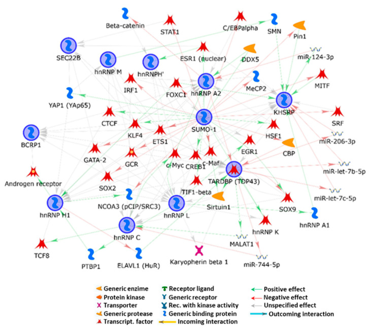
Physiologically, smooth muscle cells (SMC) and nitric oxide (NO) produced by endothelial cells strictly cooperate to maintain vasal homeostasis. In atherosclerosis, where this equilibrium is altered, molecules providing exogenous NO and able to inhibit SMC proliferation may represent valuable antiatherosclerotic agents. Searching for dual antiproliferative and NO-donor molecules, we found that furoxans significantly decreased SMC proliferation in vitro, albeit with different potencies. We therefore assessed whether this property is dependent on their thiol-induced ring opening. Indeed, while furazans (analogues unable to release NO) are not effective, furoxans' inhibitory potency parallels with the electron-attractor capacity of the group in 3 of the ring, making this effect tunable. To demonstrate whether their specific block on G1-S phase could be NO-dependent, we supplemented SMCs with furoxans and inhibitors of GMP- and/or of the polyamine pathway, which regulate NO-induced SMC proliferation, but they failed in preventing the antiproliferative effect. To find the real mechanism of this property, our proteomics studies revealed that eleven cellular proteins (with SUMO1 being central) and networks involved in cell homeostasis/proliferation are modulated by furoxans, probably by interaction with adducts generated after degradation. Altogether, thanks to their dual effect and pharmacological flexibility, furoxans may be evaluated in the future as antiatherosclerotic molecules.

Keywords: atherosclerosis; furoxans; nitric oxide; proteomics; small ubiquitin-related modifier 1; smooth muscle cell proliferation.


Conflict of interest statement

The authors declare no conflict of interest.

Figures

Scheme 1
Scheme 1
Proposed mechanism of furoxans’ NO release and of thiol bioactive adduct formation; R1 = group in position 3; R2 = group in position 4.
Figure 1
Figure 1
Effect of 3-cyano-4-phenylfuroxan (compound 12), 4-cyano-3-phenylfuroxan (compound 16), and their corresponding ineffective furazan (compound 25) on proliferation of human SMCs. Cells were seeded at a density of 7 × 10⁴/Petri dish (35 mm) and incubated with MEM supplemented with 10% FCS. Twenty-four hours later, the medium was changed to one containing 0.4% FCS to stop cell growth, and the cultures were incubated for 72 h. At this time, the medium was replaced with one containing 10% FCS in the presence or absence (control) of known concentrations of the tested compounds, and the incubation was continued for a further 72 h at 37 °C. Cell proliferation was evaluated by cell count after trypsinization of the monolayers by a Coulter Counter model Z. Values are the mean ± SD of three different experiments, each run in triplicate * p < 0.05; ** p < 0.01; *** p < 0.001 vs. control (Student’s t-test).
Figure 2
Figure 2
Linear relationship between the potency (µM) in inhibiting human SMC cell proliferation and vasodilating activity on rat aorta strips elicited by 4-Ph-furoxans with different substituents in position 3.
Figure 3
Figure 3
Effect of 3-acetoxy-4-phenylfuroxan (compound 11) vs. its corresponding ineffective furazan (compound 24) on thymidine incorporation in synchronized rat SMCs. Cells were seeded at 3 × 10⁵ cells/plate and synchronized by growing them for 120 h in a medium containing 0.4% FCS. Quiescent cells were then incubated for 20 h in fresh medium with 10% FCS, in the presence of the tested compounds. DNA synthesis was estimated by nuclear incorporation of [3H]-thymidine, incubated with cells (2 uCi /mL) for 2 h. Values are the mean ± SD of one experiment run in triplicate *** p < 0.001 vs. control (Student’s t-test).
Figure 4
Figure 4
Effect of putrescine and ODQ, alone or associated, on thymidine incorporation in synchronized rat SMC treated with furoxan 14 or SNAP. Experimental conditions are the same as in Figure 3.
Figure 5
Figure 5
Upper panel: Venn diagram representing the 11 proteins modified by furoxan 12 as documented by SILAC technique. Lower panel: Table with ratios H/L, L/H, and significance B of the 11 modified proteins as found by direct and reverse experiments conducted by SILAC technique.
Figure 6
Figure 6
MetaCore network analysis of the related molecular processes and biochemical pathways underlined by their functional cooperation in furoxans’ antiproliferative effect on SMCs. SUMO1 appears as the central hub.
Figure 7
Figure 7
Western blot and densitometric analysis of the effect of furoxan 12 on SUMO1 expression in SMCs. *** p < 0.001 vs. control (Student’s t-test).

References

    1. Raines E.W. The Extracellular Matrix Can Regulate Vascular Cell Migration, Proliferation, and Survival: Relationships to Vascular Disease. Int. J. Exp. Pathol. 2000;81:173–182. doi: 10.1046/j.1365-2613.2000.00155.x. - DOI - PMC - PubMed
    1. Williams K.J., Tabas I. The Response-to-Retention Hypothesis of Early Atherogenesis. Arterioscler. Thromb. Vasc. Biol. 1995;15:551–561. doi: 10.1161/01.ATV.15.5.551. - DOI - PMC - PubMed
    1. Borén J., Chapman M.J., Krauss R.M., Packard C.J., Bentzon J.F., Binder C.J., Daemen M.J., Demer L.L., Hegele R.A., Nicholls S.J., et al. Low-Density Lipoproteins Cause Atherosclerotic Cardiovascular Disease: Pathophysiological, Genetic, and Therapeutic Insights: A Consensus Statement from the European Atherosclerosis Society Consensus Panel. Eur. Heart J. 2020;41:2313–2330. doi: 10.1093/eurheartj/ehz962. - DOI - PMC - PubMed
    1. Björkegren J.L.M., Lusis A.J. Atherosclerosis: Recent Developments. Cell. 2022;185:1630–1645. doi: 10.1016/j.cell.2022.04.004. - DOI - PMC - PubMed
    1. Libby P. The Changing Landscape of Atherosclerosis. Nature. 2021;592:524–533. doi: 10.1038/s41586-021-03392-8. - DOI - PubMed