Review
. 2023 Jul 25;23(15):6671.
doi: 10.3390/s23156671.

Wearable Optical Fiber Sensors in Medical Monitoring Applications: A Review

Affiliations
Review

Wearable Optical Fiber Sensors in Medical Monitoring Applications: A Review

Xuhui Zhang et al. Sensors (Basel). .

Abstract

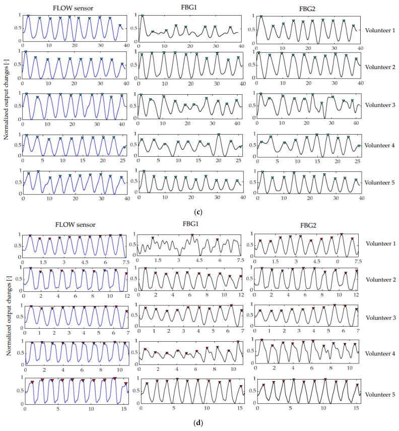
Wearable optical fiber sensors have great potential for development in medical monitoring. With the increasing demand for compactness, comfort, accuracy, and other features in new medical monitoring devices, the development of wearable optical fiber sensors is increasingly meeting these requirements. This paper reviews the latest evolution of wearable optical fiber sensors in the medical field. Three types of wearable optical fiber sensors are analyzed: wearable optical fiber sensors based on Fiber Bragg grating, wearable optical fiber sensors based on light intensity changes, and wearable optical fiber sensors based on Fabry-Perot interferometry. The innovation of wearable optical fiber sensors in respiration and joint monitoring is introduced in detail, and the main principles of three kinds of wearable optical fiber sensors are summarized. In addition, we discuss their advantages, limitations, directions to improve accuracy and the challenges they face. We also look forward to future development prospects, such as the combination of wireless networks which will change how medical services are provided. Wearable optical fiber sensors offer a viable technology for prospective continuous medical surveillance and will change future medical benefits.

Keywords: fiber Bragg grating; healthcare; optical fiber sensors; wearable sensors.


Conflict of interest statement

The authors declare no conflict of interest.

Figures

Figure 1
Figure 1
FBG encapsulated in a flexible material (a) and FBG encapsulated in dumbbell silicone (b).
Figure 2
Figure 2
(a) Schematic diagram of chest breathing monitoring (i) and correlation between FBG wavelength change and UT volume (L) (ii) [63]; (b) schematic diagram of thoracoabdominal breathing monitoring (i) and FBG volume (L) change (ii) [50]; (c) the schematic diagram of chest and abdomen respiratory monitoring (i) and the VT value calculated by smart textile are compared with the VT value collected by motion (ii) [51].
Figure 3
Figure 3
The shoulder neck monitoring structure diagram (a) and the output changes of wearable (black line) and motion capture (mobcap) systems (blue line) collected during shoulder neck up, down, left, and proper repetition (b). The flowmeter (blue line) and FBG (black line) are the signals collected by each volunteer during quiet breathing (c) and rapid breathing (d) [45].
Figure 3
Figure 3
The shoulder neck monitoring structure diagram (a) and the output changes of wearable (black line) and motion capture (mobcap) systems (blue line) collected during shoulder neck up, down, left, and proper repetition (b). The flowmeter (blue line) and FBG (black line) are the signals collected by each volunteer during quiet breathing (c) and rapid breathing (d) [45].
Figure 4
Figure 4
(a) Schematic diagram of thoracoabdominal breathing monitoring and 5 FBGs of the chest (i) and abdomen (ii) detected strain patterns under four states of standing, sitting, lying down, and running [22]; (b) schematic diagram of thoracoabdominal breathing monitoring (i) and strain detected by FBG under various postures [47] (ii).
Figure 4
Figure 4
(a) Schematic diagram of thoracoabdominal breathing monitoring and 5 FBGs of the chest (i) and abdomen (ii) detected strain patterns under four states of standing, sitting, lying down, and running [22]; (b) schematic diagram of thoracoabdominal breathing monitoring (i) and strain detected by FBG under various postures [47] (ii).
Figure 5
Figure 5
(a) Schematic diagram of respirator for respiratory monitoring (b) detection of the respiratory curve and peak wavelength drift in normal respiratory cycle (i) and slow respiratory cycle (ii) [95].
Figure 6
Figure 6
(a) Schematic diagram of elbow joint monitoring (i) and the relationship between elbow angle and Bragg wavelength displacement before and after movement (ii) [29]; (b) schematic diagram of knee joint monitoring and kind response during walking (i), enlarged image of a gait cycle (ii) [46]; (c) lumbar monitoring schematic diagram (i) and wearable output  λB, distance between L1 and L3, lumbar angle obtained according to e θ trend (ii) [36]; (d) gait monitoring diagram (i) and simultaneous response of kids and fluids during walking (ii) [47].
Figure 6
Figure 6
(a) Schematic diagram of elbow joint monitoring (i) and the relationship between elbow angle and Bragg wavelength displacement before and after movement (ii) [29]; (b) schematic diagram of knee joint monitoring and kind response during walking (i), enlarged image of a gait cycle (ii) [46]; (c) lumbar monitoring schematic diagram (i) and wearable output  λB, distance between L1 and L3, lumbar angle obtained according to e θ trend (ii) [36]; (d) gait monitoring diagram (i) and simultaneous response of kids and fluids during walking (ii) [47].
Figure 7
Figure 7
(a) Structural diagram and dynamic response of FBG sensor in the range of 0–19° (i) and 13°–35° (ii) compared with IMU [70]; (b) structure diagram (i) and angle results measured by MCP joint (upper) and PIP joint (lower), FBG sensor and IMU sensor (ii) [48]; (c) structure diagram (i) and the length of measured angle and actual angle (measured by goniometer) relative to index finger: 117 mm, 97 mm, 77 mm. Measure the linearity of the MCP joint angle according to the algorithm and finger length (ii) [42].
Figure 7
Figure 7
(a) Structural diagram and dynamic response of FBG sensor in the range of 0–19° (i) and 13°–35° (ii) compared with IMU [70]; (b) structure diagram (i) and angle results measured by MCP joint (upper) and PIP joint (lower), FBG sensor and IMU sensor (ii) [48]; (c) structure diagram (i) and the length of measured angle and actual angle (measured by goniometer) relative to index finger: 117 mm, 97 mm, 77 mm. Measure the linearity of the MCP joint angle according to the algorithm and finger length (ii) [42].
Figure 8
Figure 8
(a) the principle of changing light intensity is changing the distance between two optical fibers and schematic diagram of the process of light passing through an optical fiber (iiii); (b) changing the light intensity by changing the direct distance between the optical fiber and the mirror and schematic diagram of the process of light passing through an optical fiber (iiii); (c) changing the light intensity principle by macro bending of optical fibers and schematic diagram of the process of light passing through an optical fiber(i,ii).
Figure 9
Figure 9
(a) Finger joint monitoring structure diagram (i) and sensor response to angular displacement change of finger joint (ii) [114]; (b) elbows joint monitoring structure diagram (i) and joint angle measurement results (ii). The blue line represents the potentiometer and the red line represents POF [65]; (c) wrist monitoring structure diagram (i) and measurement response of wrist flexion, extension, and abduction adduction motion (ii) [115]; (d) elbow joint monitoring chart (i) and average optical response of optical fiber during elbow bending of art (ii) [116]; (e) elbow and wrist joint monitoring chart (i) and output voltage increasing with carpometacarpal flexion angle (ii) [33].
Figure 9
Figure 9
(a) Finger joint monitoring structure diagram (i) and sensor response to angular displacement change of finger joint (ii) [114]; (b) elbows joint monitoring structure diagram (i) and joint angle measurement results (ii). The blue line represents the potentiometer and the red line represents POF [65]; (c) wrist monitoring structure diagram (i) and measurement response of wrist flexion, extension, and abduction adduction motion (ii) [115]; (d) elbow joint monitoring chart (i) and average optical response of optical fiber during elbow bending of art (ii) [116]; (e) elbow and wrist joint monitoring chart (i) and output voltage increasing with carpometacarpal flexion angle (ii) [33].
Figure 10
Figure 10
(a) Structure diagram of respiratory monitoring (i) and function of optical output power (1 cm in diameter) of respiratory sensor and elongation of respiratory sensor (ii) [110]; (b) respiratory monitoring structure diagram (i) and power output of optical fiber respiratory sensors with different bending times and different bending diameters (ii) [12]; (c) the time domain waveforms of respiratory monitoring structure diagram (i) and (the first three columns) original signals at rest, walking and running, respectively. (Middle three columns) time domain waveform of the respiratory signal after signal and processing. (Last three columns) frequency domain corresponding to respiratory signals ((ii) [24]).
Figure 10
Figure 10
(a) Structure diagram of respiratory monitoring (i) and function of optical output power (1 cm in diameter) of respiratory sensor and elongation of respiratory sensor (ii) [110]; (b) respiratory monitoring structure diagram (i) and power output of optical fiber respiratory sensors with different bending times and different bending diameters (ii) [12]; (c) the time domain waveforms of respiratory monitoring structure diagram (i) and (the first three columns) original signals at rest, walking and running, respectively. (Middle three columns) time domain waveform of the respiratory signal after signal and processing. (Last three columns) frequency domain corresponding to respiratory signals ((ii) [24]).
Figure 11
Figure 11
(a) Gait-assist structure diagram (i) and POF curvature response of knee flexion and extension cycle of modular exoskeleton (above), POF insole for functional electrical stimulation (FES) assisted-gait phase detection (below) (ii) [23]; (b) gait help structure diagram (i) and ground reaction force (GRF) and plantar pressure of female subjects weighing 46kg (above). The solid line is the average GRF, and the shadow curve is the standard deviation of 5 cycles; (below) GRF and plantar pressure measurements are standardized for all 20 participants. Solid lines represent the mean GRF and shaded curves represent the standard deviation (ii) [60].
Figure 11
Figure 11
(a) Gait-assist structure diagram (i) and POF curvature response of knee flexion and extension cycle of modular exoskeleton (above), POF insole for functional electrical stimulation (FES) assisted-gait phase detection (below) (ii) [23]; (b) gait help structure diagram (i) and ground reaction force (GRF) and plantar pressure of female subjects weighing 46kg (above). The solid line is the average GRF, and the shadow curve is the standard deviation of 5 cycles; (below) GRF and plantar pressure measurements are standardized for all 20 participants. Solid lines represent the mean GRF and shaded curves represent the standard deviation (ii) [60].
Figure 12
Figure 12
Wearable ankle monitoring device based on FPI (a) and optical power variation with angular displacement. Points are experimental data, and straight lines correspond to linear fitting (b) [23].

References

    1. Leal A.G., Diaz C.A.R., Avellar L.M., Pontes M.J., Marques C., Frizera A. Polymer Optical Fiber Sensors in Healthcare Applications: A Comprehensive Review. Sensors. 2019;19:3156. doi: 10.3390/s19143156. - DOI - PMC - PubMed
    1. Gong Z., Xiang Z., OuYang X., Zhang J., Lau N., Zhou J., Chan C.C. Wearable Fiber Optic Technology Based on Smart Textile: A Review. Materials. 2019;12:3311. doi: 10.3390/ma12203311. - DOI - PMC - PubMed
    1. Umapathi R., Park B., Sonwal S., Rani G.M., Cho Y., Huh Y.S. Advances in optical-sensing strategies for the on-site detection of pesticides in agricultural foods. Trends Food Sci. Technol. 2022;119:69–89. doi: 10.1016/j.tifs.2021.11.018. - DOI
    1. Sareh S., Noh Y. Low Profile Stretch Sensor for Soft Wearable Robotics; Proceedings of the 1st IEEE-RAS International Conference on Soft Robotics (RoboSoft); Livorno, Italy. 24–28 April 2018; Piscataway Township, NJ, USA: IEEE; 2018. pp. 479–484.
    1. Li H., Yang H., Li E., Liu Z., Wei K. Wearable sensors in intelligent clothing for measuring human body temperature based on optical fiber Bragg grating. Opt. Express. 2012;20:11740–11752. doi: 10.1364/OE.20.011740. - DOI - PubMed

LinkOut - more resources