. 2024 May 2;63(5):1466-1473.
doi: 10.1093/rheumatology/kead400.

Autoantibodies against the melanoma differentiation-associated protein 5 in patients with dermatomyositis target the helicase domains

Affiliations

Autoantibodies against the melanoma differentiation-associated protein 5 in patients with dermatomyositis target the helicase domains

Eveline Van Gompel et al. Rheumatology (Oxford). .

Abstract

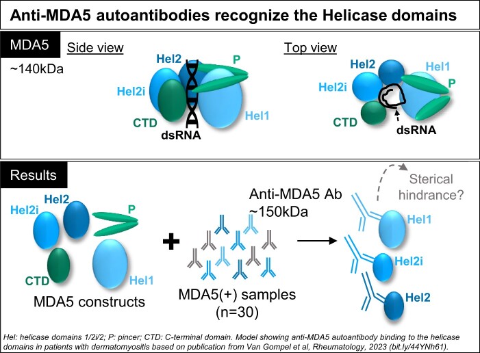
Objectives: Clinical observations in patients with dermatomyositis (DM) and autoantibodies against the melanoma differentiation-associated protein 5 (MDA5) suggest that the autoantibodies contribute to the pathogenesis of MDA5(+) DM. To gain insight into the role of the anti-MDA5 autoantibodies, we aimed to identify their binding sites on the different domains of the MDA5 protein.

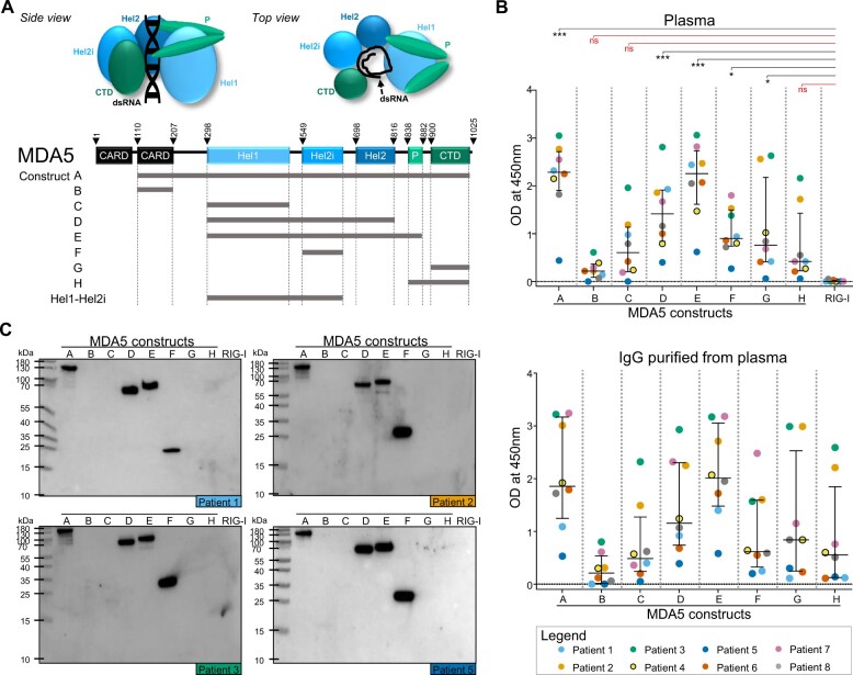
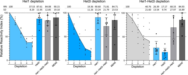
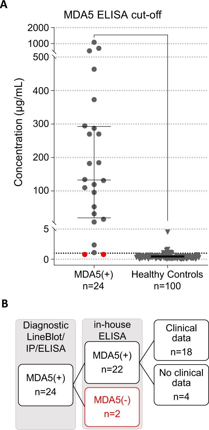
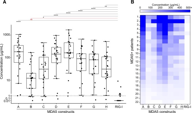
Methods: We developed an in-house ELISA to assess the reactivity against the MDA5 domains (conformational epitopes) in plasma (n = 8) and serum (n = 24) samples from MDA5(+) patients with varying clinical manifestations and disease outcomes. The reactivities were also assessed using western blot (linearized epitopes). An ELISA-based depletion assay was developed to assess cross-reactivity among the different MDA5 domains.

Results: All eight plasma samples consistently showed reactivity towards conformational and linearized epitopes on the helicase domains of the MDA5 protein. The ELISA-based depletion assay suggests that anti-MDA5 autoantibodies specifically target each of the three helicase domains. Twenty-two of the 24 serum samples showed reactivity in the in-house ELISA and all 22 displayed reactivity towards the helicase domains of the MDA5 protein.

Conclusions: Our data revealed that the main immunogenic targets of anti-MDA5 autoantibodies from MDA5(+) patients are the helicase domains. Considering that the helicase domains are responsible for the enzymatic activity and subsequent triggering of an inflammatory response, our findings suggest that binding of anti-MDA5 autoantibodies could alter the canonical activity of the MDA5 protein and potentially affect the downstream induction of a pro-inflammatory cascade.

Keywords: DM; IFIH1; IFN-induced helicase; MDA5; autoantibodies; interstitial; lung diseases; melanoma differentiation-associated protein 5.


Figures

None
Graphical abstract
Figure 1.
Figure 1.
MDA5(+) samples from plasma exchange (PLEX) show reactivity to helicase-bearing MDA5 constructs. (A) Schematic representation of the MDA5 protein (side and top view) and the MDA5 protein constructs. (B) ELISA to assess the reactivity of MDA5(+) samples from PLEX (n = 8, upper panel) and the corresponding purified IgG (n = 8, lower panel) against conformational epitopes on the MDA5 constructs A–H and the negative control (RIG-I). (C) Western blot analysis to assess the reactivity of purified IgGs against linearized epitopes on the MDA5 constructs A–H and RIG-I (n = 4 representative blots). In (B), statistical analysis was performed using Kruskal–Wallis test with Dunn’s correction: *P-value < 0.05, ***P-value < 0.001, nsP-value > 0.05. Dots represent individual subjects, lines/bars represent median (± interquartile range). MDA5: melanoma differentiation–associated protein 5; OD: optical density; CARD: N-terminal caspase activation and recruitment domains; Hel: helicase domains 1/2i/2; P: pincer; CTD: C-terminal domain; RIG-I: retinoic acid–inducible protein I
Figure 2.
Figure 2.
Depletion ELISA shows anti-MDA5 autoantibodies specifically target the helicase domains of the MDA5 protein. Relative Reactivity Index (RRI, residual reactivity as a fraction of the original reactivity, in %) of IgG purified from MDA5(+) plasma samples (n = 8) during and after depletion in ELISA. Dots represent individual subjects, lines/bars represent mean (± s.d.). MDA5: melanoma differentiation–associated protein 5; Hel: helicase domains 1/2i/2
Figure 3.
Figure 3.
Receiver operating characteristic curve analysis to determine the cut-off for positivity of the in-house MDA5 ELISA. (A) The cut-off for reactivity against MDA5 construct A in the in-house MDA5 ELISA was determined based on subjects diagnosed with MDA5(+) DM (n = 24) and healthy control subjects (n = 100) using receiver operating characteristic analysis and set at 1.07 µg/ml (dotted line). Red dots indicate samples below cut-off. Dots represent individual patients, lines/bars represent median (± interquartile range). (B) Flow diagram of in-/excluded patients with MDA5(+) DM. MDA5: melanoma differentiation–associated protein 5; IP: immunoprecipitation
Figure 4.
Figure 4.
MDA5(+) sera show reactivity to multiple MDA5 constructs, with the main reactivity directed towards helicase-bearing constructs. (A) Reactivity profile of MDA5(+) serum samples (n = 22) in in-house ELISA, reported as anti-MDA5 autoantibody levels calculated based on a standard curve consisting of a serial dilution of purified anti-MDA5 autoantibodies. The black dotted line represents the cut-off for reactivity against construct A. Statistical analysis was performed using Kruskal–Wallis test with Dunn’s correction: *P-value < 0.05, **P-value < 0.01, ****P-value < 0.0001, nsP-value > 0.05. Dots represent individual patients, lines/bars represent median (± interquartile range). (B) Heatmap showing the reactivity profiles in individual patient samples, ranked from highest to lowest reactivity against construct A. MDA5: melanoma differentiation–associated protein 5

References

    1. Betteridge Z, McHugh N.. Myositis-specific autoantibodies: an important tool to support diagnosis of myositis. J Intern Med 2016;280:8–23. - PubMed
    1. McHugh NJ, Tansley SL.. Autoantibodies in myositis. Nat Rev Rheumatol 2018;14:290–302. - PubMed
    1. Aggarwal R, Oddis CV, Goudeau D. et al. Autoantibody levels in myositis patients correlate with clinical response during B cell depletion with rituximab. Rheumatology (Oxford) 2016;55:991–9. - PMC - PubMed
    1. Moghadam-Kia S, Aggarwal R, Oddis CV.. Biologics for idiopathic inflammatory myopathies. Curr Opin Rheumatol 2017;29:645–51. - PubMed
    1. Aggarwal R, Charles-Schoeman C, Schessl J. et al. ; ProDERM Trial Group. Trial of intravenous immune globulin in dermatomyositis. N Engl J Med 2022;387:1264–78. - PubMed

Publication types

Substances