. 2023 Aug 11;4(8):554-579.e9.
doi: 10.1016/j.medj.2023.07.004.

Organoid co-culture model of the human endometrium in a fully synthetic extracellular matrix enables the study of epithelial-stromal crosstalk

Affiliations

Organoid co-culture model of the human endometrium in a fully synthetic extracellular matrix enables the study of epithelial-stromal crosstalk

Juan S Gnecco et al. Med. .

Abstract

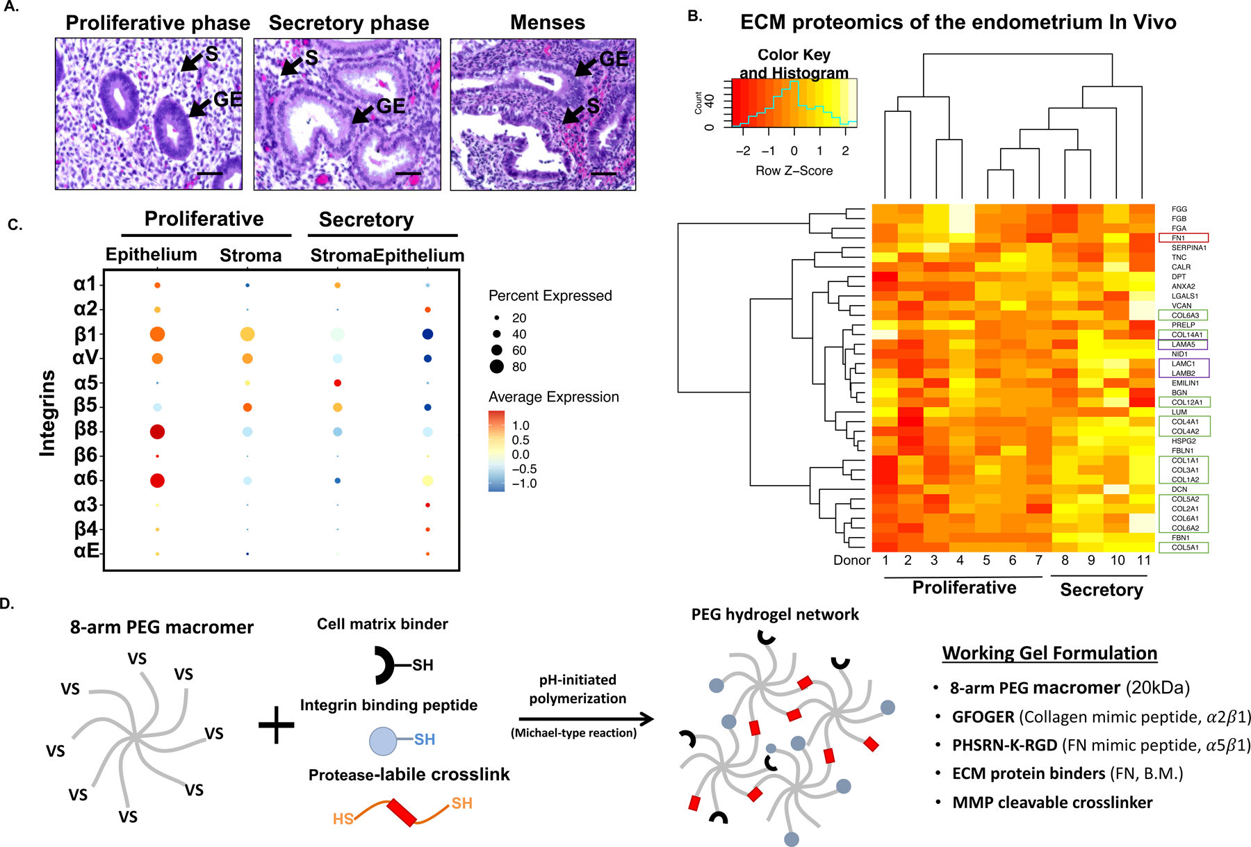
Background: The human endometrium undergoes recurring cycles of growth, differentiation, and breakdown in response to sex hormones. Dysregulation of epithelial-stromal communication during hormone-mediated signaling may be linked to myriad gynecological disorders for which treatments remain inadequate. Here, we describe a completely defined, synthetic extracellular matrix that enables co-culture of human endometrial epithelial and stromal cells in a manner that captures healthy and disease states across a simulated menstrual cycle.

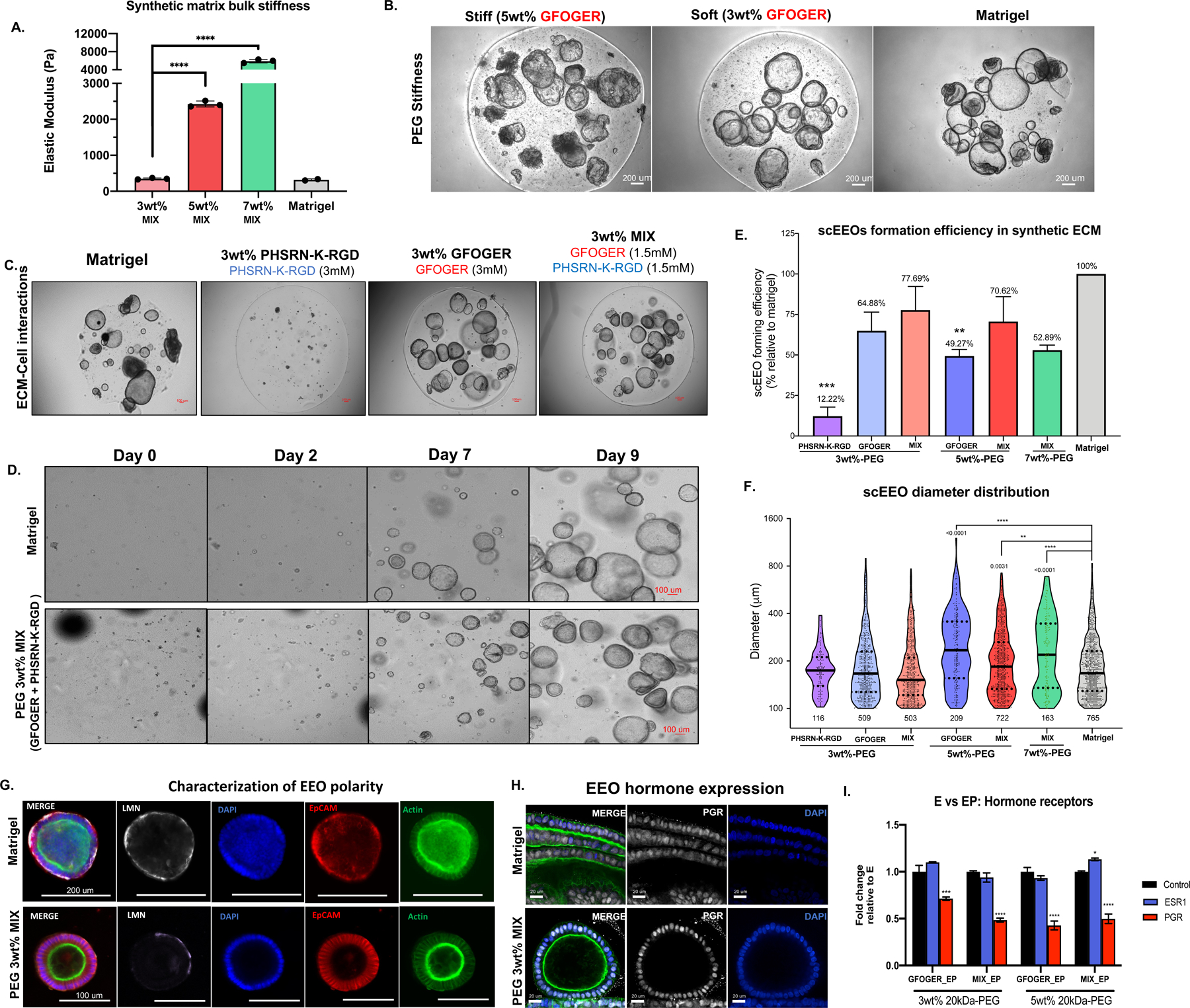
Methods: We parsed cycle-dependent endometrial integrin expression and matrix composition to define candidate cell-matrix interaction cues for inclusion in a polyethylene glycol (PEG)-based hydrogel crosslinked with matrix metalloproteinase-labile peptides. We semi-empirically screened a parameter space of biophysical and molecular features representative of the endometrium to define compositions suitable for hormone-driven expansion and differentiation of epithelial organoids, stromal cells, and co-cultures of the two cell types.

Findings: Each cell type exhibited characteristic morphological and molecular responses to hormone changes when co-encapsulated in hydrogels tuned to a stiffness regime similar to the native tissue and functionalized with a collagen-derived adhesion peptide (GFOGER) and a fibronectin-derived peptide (PHSRN-K-RGD). Analysis of cell-cell crosstalk during interleukin 1B (IL1B)-induced inflammation revealed dysregulation of epithelial proliferation mediated by stromal cells.

Conclusions: Altogether, we demonstrate the development of a fully synthetic matrix to sustain the dynamic changes of the endometrial microenvironment and support its applications to understand menstrual health and endometriotic diseases.

Funding: This work was supported by The John and Karine Begg Foundation, the Manton Foundation, and NIH U01 (EB029132).

Keywords: Foundational research.


Conflict of interest statement

Declaration of interests L.G.G. and V.H.G. have a patent application pending related to the hydrogel system.

Figures

Figure 1.
Figure 1.. A tissue-inspired multi-omic approach elucidates the design parameters to engineer a fully synthetic PEG hydrogel for the endometrium.
(A) Representative histological hematoxylin and eosin (H&E) stained samples of the menstrual cycle phases. Arrows (▲) indicate stromal stromal fibroblasts (S) and the glandular epithelium (GE). (B) Heatmap of proteomic analysis of the human endometrium showing ECM proteins (matrisome) across the menstrual cycle (n=11 donors). Box colors indicated ECM protein associations: green corresponds to collagens; red to fibronectin; and purple to laminins. (C) Dot plot analysis of specific integrins expression from single cell RNA-seq data (n = 6 donors, 6,659 cells). Examination of stromal and epithelial component integrin expression across the menstrual cycle (proliferative and secretory phases). Circle size and color indicate P-value and variation about the means of the average expression value for each integrin chain in all cells (D) Schematic of the overall strategy to design and fabricate a synthetic ECM for the endometrium. 8-arm 20kDa PEG macromers are functionalized with cell matrix binders (FN, B.M.), integrin adhesion peptides, and polymerized with a protease-labile crosslinker. Descriptive outline of the key peptides utilized in the synthetic ECM formulation.
Figure 2.
Figure 2.. Dual integrin ligand functionalization in softer synthetic matrix regimes enhance EEOs generation.
(A) Biomechanical properties (elastic moduli, Pa, N=3 gels) of synthetic hydrogel formulations, where “soft” = 3wt% (~300 Pa), “stiff” = 5wt% (~2kPa) and “stiffest” = 7wt% (~6kPa). (B) Representative images of scEEO and morphology in stiff (5wt%) in soft (3wt%) PEG hydrogels functionalized with only GFOGER (3mM) compared to Matrigel. (C) Day-15 images of hydrogel formulations comparing adhesion peptides reveals that GFOGER is necessary to generate scEEOs. Dual-adhesion-peptide functionalization with GFOGER (1.5 mM) and PHSRN-K-RGD (1.5 mM) is sufficient for robust scEEO generation. (D) Time-lapse images of 10-day cultures of EEO derived from single cells in synthetic hydrogels (PEG 3wt%-MIX). (E) Quantification of the scEEO formation efficiency in synthetic hydrogels relative to Matrigel (N=8). (F) Quantification of the total lumenized EEO (>100 um diameter) count and EEO diameter distribution after 10 days in culture. (G) Characterization of EEO morphology and polarity of EEOs generated in synthetic ECM (3wt%-MIX) compared to Matrigel via immunofluorescent (IF) analysis of ECM deposition for laminin (LMN) and F-actin localization after 14 days of culture in soft (3%) PEG MIX hydrogel and in Matrigel. (H-I) Characterization of protein and transcriptomic expression of hormone receptors in EEOs generated in synthetic hydrogels. (H) Representative images of PGR staining countered stained with F-actin (green) and DAPI (nuclei) and (I) qPCR analysis of mRNA expression of PGR and ESR1 in EEOs treated with either E2 (E) or E2 + MPA (EP) for 14 days. Analysis compares ESR1, PGR values relative to matching E treatment groups (control). Significance is indicated as *p<0.05, **p<0.01, ***p<0.001.
Figure 3.
Figure 3.. Development and characterization of a synthetic endometrial stromal cell culture.
(A) Schematic of the workflow for culturing ESCs in 3D using a synthetic hydrogel. (B) Maximum intensity projection images of F-actin (green) reveal ESC remodeling (cell elongation and dispersion) at day 7 of culture in stromal (serum containing) media impacted primarily by hydrogel stiffness (20X magnification) as quantified in (C). (D) Time-lapse images of embedded ESCs in synthetic matrix (PEG) compared to the natural-derivedhydrogels Collagen I (~4 mg/mL) and Matrigel (8 mg/mL) allow for long-term stable cultures. (E) Brightfield images of stromal decidualization morphology in synthetic ECMs as a function of matrix stiffness (3wt%, 5wt% and 7wt% MIX) after 14 days of EP treatment and in standard ESC media or EEO medium (see Methods). (F) Biochemical measurement of prolactin (PRL) in the spent media (EEO media) from 15-day cultures in (E) compared to standard 2D polystyrene cultures (N=4). (G) Represenatitive fluorescent staining of DNA synthesis (EdU incorporation for 24 hrs hrs prior to fixation at day 15 as a function of simulated cycle phase and inflammation cue, IL1B (H) Quantification of mean EdU+ staining from cultures shown in (G) shows IL1B suppresses DNA synthesis of ESCs (n=3) (I) Quantification of apoptosis (cleaved caspase-3+) in ESC cells stimulated with homrone and IL1B treatment groups at day 15 of culture relative to total nuclei count (N=3) (J) Inflammatory challenge with IL1B significantly suppressed PRL secretion in ESC cultures (E+ = E + IL1B, PE+= PE+IL1B, W = hormone withdrawal) (N=6). (K) Vimentin stain (red) of ESC morphology cultured in synthetic and natural hydrogels. IL1B (1ng/mL) treated ESC in synthetic hydrogel provided as a control for inflamed morphology after 6 days. Significance is indicated as *p<0.05, **p<0.01, ***p<0.001. Scale bars are 50 mm, unless otherwise noted.
Figure 4.
Figure 4.. Establishment of a co-culture model recapitulates the molecular signature of the human menstrual cycle in vitro.
(A) Schematic and representative images of the co-cultures. EEOs (10 intact organoids/µL) and ESC (10k cells/µL) are expanded separately and embedded in the synthetic ECM, cultured as 3 µl droplets and maintained for 15 days of culture. A dual-adhesion peptide synthetic ECM (3wt% 20kDa-PEG) functionalized with GFOGER (1.5 mM) and PHSRN-K-RGD (1.5mM) supports the establishment of the EEOs and ESC co-culture model. (B) Representative 3D images of day-15 endometrial EEOs (EpCAM = green) and ESC (Vimentin = red) in the co-cultures (scale = 100 µm). Images acquired from Video S3 in Data File 3. (C) Schematic and timeframe of the idealized 28-day menstrual cycle in vivo. (D) Experimental design for the validation of the co-culture endometrial model. Treatment groups are designed to mimic the hormonal changes in the phases of the menstrual cycle, including a pharmacologic induction of the menstruation using a PGR antagonist (RU-486, 10 µM). ‘Inflamed’ groups were co-treated with IL1B (1ng/mL) throughout the length of the experiment. (E-G) Transcriptomic (bulk RNAseq) analysis of the co-culture models (n= 7). (E) Gene ontology (GO) analysis of the top significantly enriched GO terms that are downregulated or upregulated in response to progestin treatment. Cocultures treated with E2 + MPA (“PE”) have gene ontology (GO) expression profiles that align with the secretory phase of the menstrual cycle. GSEA analysis of hallmark pathway gene sets show pathway changes consistent with corresponding phases of the menstrual cycle. All images and transcriptomic analysis were performed on day-15 co-cultures in the synthetic 3wt% 20kDa-PEG-Mix hydrogels (N=8). Data is shown with the −log of their P values.
Figure 5.
Figure 5.. Functional validation of the co-culture model recapitulates morphologic and biochemical cycle-dependent reproductive processes.
(A) Representative histological (IF) characterization of the proliferative and secretory phases of the menstrual cycle in vivo reveals morphologic and biochemical changes. Vimentin (red), F-Actin (Green), DAPI (blue). (B) Characterization of the epithelial morphology in co-cultures at day 15 of culture by IF reveals glandular maturation and invagination of EEOs in response to progestin treatment groups. (C) IF staining of PAEP (glycodelin) in co-cultures recapitulates the epithelial secretory phenotype. (D) Stromal decidualization was assessed by measuring prolactin secretion in the medium across 15 days of culture. PRL secretion is induced by PE treatment but suppressed in menses treatment group. (E) PGR staining in the co-cultures at day 15 of treatment. (F) Temporal profiles of decidualization normalized to fold change relative to day 0 were performed by measuring spent media (N=12) and can be suppressed by IL1B treatment (1 ng/mL, E+, PE+). (G-H) Cleaved caspase-3 (Casp-3) IF in the co-cultures as a marker of apoptosis (G) and quantified in as positive staining per organoid area (H). All images are of day-15 co-cultures in the synthetic ECM 3wt% 20kDa-PEG-Mix hydrogels. Significance is indicated as *p<0.05, **p<0.01, ***p<0.001.
Figure 6.
Figure 6.. Functional analysis of inflammatory cue (IL1B) repsonses in co-cultures in synthetic hydrogel reveals the intitiation of the endometriotic phenotype mediated by cell-cell communication.
(A) 3D maximum intensity projections of PGR expresison in E2-only treated co-cultures in response to IL1B stimulation (B-C) IL1B stimulation (1 ng/mL) induces morphological changes in EEOs. Representative IF images (B) and quantification (C) of epithelial height (distance from base to apical edge of the epithelium, N=3). (D) Schematic of donor matched EEO monoculture and co-culture in IL1B treatment groups. (E) Represenative images of time-lapse co-cultures stimulated with IL1B reveals enlarged EEO diameter by day 7 of culture. Quantication of the mean fold-change of EEO diameter across 15-days of cultures from daily time-lapse images in co-cultures (black) and monocultures (red) across treatment conditions (E, PE, W) and IL1B-treated (E+, PE+) groups. Fold-change denotes the mean increase in diameter relative to day 1 of culture. Analysis compares the mean EEO growth rate between monoculture vs coculture (n=4; E + IL-1 p=0.0076; PE + IL1B p=0.0026). (G) Immunostaining analysis of DNA synthesis in co-cultures and EEO monocultures by EdU incorporation in response to hormones and IL1B treatment (N=3). (H-I) Quantification of proliferative profiles calculated as the number of EdU+ cells per organoid area (mm2) in (H) organoid monocultures and (I) co-cultures. (J) Working model of the cellular mechanisms driving epithelial and stromal communication in the endometrium under physiologic (hormones) and pathogenic (IL1B-treated) conditions. All images are of day-15 co-cultures in the synthetic ECM 3wt% 20kDa-PEG-MIX hydrogels. Significance is indicated as *p<0.05, **p<0.01, ***p<0.001.

Comment in

Similar articles

Cited by

References

    1. Aplin JD (1989). Cellular Biochemistry of the Endometrium. In Biology of the Uterus (Karger Publishers), pp. 89–129. 10.1007/978-1-4684-5589-2_6. - DOI
    1. Aplin JD, and Ruane PT (2017). Embryo-epithelium interactions during implantation at a glance. J. Cell Sci 130, 15–22. 10.1242/jcs.175943. - DOI - PubMed
    1. Loy SLY and S T (2005). Normal cycling Endometrium:Molecular, Cellular, and Histologic Perspectives. In Endometriosis in Clinical Practice, pp. 1–29.
    1. Noyes RW, Hertig AT, and Rock J (1975). Dating the endometrial biopsy. Am. J. Obstet. Gynecol 122, 262–263. 10.1016/S0002-9378(16)33500-1. - DOI - PubMed
    1. Roy A, and Matzuk MM (2011). Reproductive tract function and dysfunction in women. Nat. Rev. Endocrinol 7, 517–525. 10.1038/nrendo.2011.79. - DOI - PubMed

Publication types

LinkOut - more resources