. 2023 Oct 5;31(10):1233-1246.e5.
doi: 10.1016/j.str.2023.07.009. Epub 2023 Aug 11.

Structural basis of microtubule depolymerization by the kinesin-like activity of HIV-1 Rev

Affiliations

Structural basis of microtubule depolymerization by the kinesin-like activity of HIV-1 Rev

Elif Eren et al. Structure. .

Abstract

HIV-1 Rev is an essential regulatory protein that transports unspliced and partially spliced viral mRNAs from the nucleus to the cytoplasm for the expression of viral structural proteins. During its nucleocytoplasmic shuttling, Rev interacts with several host proteins to use the cellular machinery for the advantage of the virus. Here, we report the 3.5 Å cryo-EM structure of a 4.8 MDa Rev-tubulin ring complex. Our structure shows that Rev's arginine-rich motif (ARM) binds to both the acidic surfaces and the C-terminal tails of α/β-tubulin. The Rev-tubulin interaction is functionally homologous to that of kinesin-13, potently destabilizing microtubules at sub-stoichiometric levels. Expression of Rev in astrocytes and HeLa cells shows that it can modulate the microtubule cytoskeleton within the cellular environment. These results show a previously undefined regulatory role of Rev.

Keywords: CTT; HIV-1; Microtubules; Rev; Rings; Tubulin.


Conflict of interest statement

Declaration of interests The authors declare that they have no conflicts of interest with the contents of this article.

Figures
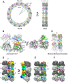
Figure 1.
Figure 1.. Structure of the Rev-tubulin complex.
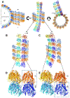
(A) The Rev-tubulin ring-complex viewed from front (left) and side (right) with β-tubulin (dark gray), α-tubulin (light gray), and the Rev monomers in different colors, all depicted as ribbons. (B) The asymmetric unit (ASU), as viewed from the center of the ring with the plus-end up, relative to a microtubule (left), and the ASU viewed towards the plus-end (right). Rev monomers: E, dark green; F, light green; G, orange; H, yellow; I, dark blue; and J, cyan. (C) The minimal biological assembly (MBA), consisting of α-β-tubulin heterodimers, as viewed from the center of the ring with the plus-end up, with the subunits colored as above. (D) The MBA rendered as a surface as viewed from the center of the ring (left) and viewed from the side of the primary ring (right). Rev and tubulin subunits are labeled uniquely and the chain IDs match the deposited structure (PDB ID: 7U0F). (E) The MBA with the Rev monomers removed and their contact areas on tubulin colored accordingly, as viewed form the center of the ring. Areas I, II, III indicated (see text). (F) The MBA with the contact areas of the Rev monomers on the CTTs of the adjacent tubulin subunits colored accordingly.
Figure 2.
Figure 2.. Charge and hydrophobic interactions between Rev and tubulin.
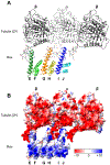
(A) Surface-charge representation of the tubulin and Rev components of the ring-complex; Red, negative; blue, positive; white neutral. The color ranges from −10 (red) to +10 (blue) kcal/(mol·e) at 298 °K. The tubulin (left), as viewed from the center of the ring. The Rev assembly (right) rotated to show the face interacting with tubulin. (B) Surface-hydrophobicity representation of the tubulin and Rev components of the ring-complex; blue, hydrophilic; orange, hydrophobic; and white neutral (Kyte-Doolittle hydropathy scale). The Rev-Rev interaction is hydrophobic, the Rev-tubulin interaction is charge-mediated.
Figure 3.
Figure 3.. Rev interactions with the secondary tubulin ring.
(A) Interaction of Rev monomers in the ASU with the tubulin heterodimer and the adjacent β-tubulin C-termini. β-tubulin is colored in dark gray, and α-tubulin is colored in light gray. ASU Rev monomers are shown in different colors (as coded in Figure 1). (B) Surface-charge representation of the tubulin (in 2ry ring) and Rev; Red, negative; blue, positive; white neutral. The color ranges from −10 (red) to +10 (blue) kcal/(mol·e) at 298 °K
Figure 4.
Figure 4.. Effect of CTT removal on Rev-tubulin ring formation.
(A) Representative negative stain image of Taxol-tubulin + Rev (B) Subtilisin treated Taxol-tubulin + Rev (C) Subtilisin treated tubulin + Rev. (D) Close-up view of stacked single rings (from panel C) formed by Subtilisin treated tubulin + Rev.
Figure 5.
Figure 5.. Comparison of Rev and Kinesin-13 interactions with tubulin.
(A) The MBA, as viewed from the side of the primary ring; α-tubulin, light gray; β-tubulin, dark gray. Rev monomers are colored as in Figure 1. Three areas of contact with the heterodimer (I, II, and III) are indicated. (B) Kin-13 KLP10A (PDB: 6B0C) bound to tubulin as shown from the same point of view as in (A). KLP10A is shown in magenta and the three contact areas are indicated. (C) KLP10A-tubulin aligned to Rev-tubulin. Tubulin contacts made by 6 Rev monomers are similar to contacts made by a KLP10A monomer. (D) Close-up view of Area III. KLP10A L2 contacts on α-tubulin are shown in orange and on β-tubulin are shown in brown. Conserved L2 loops KVD are shown. Rev contacts on α-tubulin are shown in green. Residues that interact both with KLP10A and Rev are shown in blue.
Figure 6.
Figure 6.. Structural relationship between rings and microtubules.
(A) The Rev-tubulin ring-complex aligned to the inner microtubule of the KLP10A-tubulin complex (PDB: 6B0C), as viewed in three orthogonal orientations. Left, the first (I) quadrant of the ring complex as positioned by alignment of a primary β-subunit with a β-subunit of the microtubule (representing a complex formed at the plus-end), and the fourth (IV) quadrant of the ring complex as positioned by alignment of a primary α-subunit with an α-subunit of the microtubule (representing a complex formed at the minus-end). Middle, the model as viewed from the center of the ring. Right, the model as viewed towards the microtubule plus-end. Microtubule α- and β-tubulin are shown in orange and blue, respectively. Primary ring α- and β-tubulin, red orange and dark blue, respectively. Secondary ring α- and β-tubulin, yellow and cyan, respectively. (B) View similar to (A, middle) but turned slightly to show clashes with the protofilament to the left. Boxed area is enlarged in (D). (C) As in (B) but turned slightly to show the subunits oriented away from the protofilament to the right. Boxed area is enlarged in (E). (D) The four tubulin subunits of the ASU, and four tubulin subunits from the microtubule lattice, with the primary α-subunit and the microtubule α-subunit aligned (boxed). (E) As in (D) but with the primary β-subunit and the microtubule β-subunit aligned (boxed).
Figure 7.
Figure 7.. Conformational changes in tubulin subunits lead to loosening of lateral interactions.
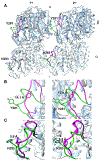
(A) Alignment of a straight microtubule (PDB: 6EVY) α- and β-tubulin heterodimers with Rev-tubulin ring complex α- and β-heterodimers for both the primary and secondary rings. The two adjacent heterodimers are viewed from the outside (the luminal side of a microtubule). M-loops of straight microtubule are shown in green and Rev-tubulin are shown in magenta. The sidechains of α-H283 and β-Y281 are shown. (B) Close-up views of Rev-primary β-tubulin M-loop, left; and Rev-secondary β-tubulin M-loop, right aligned to straight microtubule. (C) Close-up views of Rev-primary α-tubulin M-loop, left; and Rev-secondary α-tubulin M-loop, right aligned to straight microtubule. Distances between straight and curved (ring) β-Y281 and α-H283 are shown. Whereas in a microtubule, where the M-loops on both the α- and β-subunits are oriented towards the adjacent subunits, in the Rev-tubulin ring-complex they are oriented away, in both the primary and secondary rings, suggesting a weakening of the lateral interactions.
Figure 8.
Figure 8.. Confocal microscopy images depicting tubulin depolymerization.
Tubulin depolymerization in astrocytes (A) and HeLa (B) cells. Top panels show control cells, bottom panels show Rev-expressing cells. From left to right: merged, α-tubulin (magenta) detected by antibodies against α-tubulin, acetylated α-tubulin (cyan) detected by antibodies against α-tubulin K40Ac antibodies, Rev (green) detected by anti-FLAG M2 antibodies and nuclei stained with DAPI. See materials and methods section for additional details. Scale bars, 10 μm.
Figure 9.
Figure 9.. Comparison of tubulin depolymerization by nocodazole and Rev.
Tubulin depolymerization in astrocytes. Top panel shows control cells, middle panel shows Rev transfected cells and bottom panel shows 10 μM nocodazole treated cells. From left to right: merged, α-tubulin (magenta) detected by antibodies against α-tubulin, acetylated α-tubulin (cyan) detected by antibodies against α-tubulin K40Ac antibodies, Rev (green) detected by anti-FLAG M2 antibodies and nuclei stained with DAPI. Scale bars, 25 μm.

Similar articles

References

    1. Goodson HV, and Jonasson EM (2018). Microtubules and Microtubule-Associated Proteins. Cold Spring Harb Perspect Biol 10. 10.1101/cshperspect.a022608. - DOI - PMC - PubMed
    1. Cote RH, and Borisy GG (1981). Head-to-tail polymerization of microtubules in vitro. J Mol Biol 150, 577–599. 10.1016/0022-2836(81)90382-x. - DOI - PubMed
    1. Mitchison TJ (1993). Localization of an exchangeable GTP binding site at the plus end of microtubules. Science 261, 1044–1047. 10.1126/science.8102497. - DOI - PubMed
    1. Kaverina I, and Straube A (2011). Regulation of cell migration by dynamic microtubules. Semin Cell Dev Biol 22, 968–974. 10.1016/j.semcdb.2011.09.017. - DOI - PMC - PubMed
    1. Li R, and Gundersen GG (2008). Beyond polymer polarity: how the cytoskeleton builds a polarized cell. Nat Rev Mol Cell Biol 9, 860–873. 10.1038/nrm2522. - DOI - PubMed

Publication types

LinkOut - more resources