. 2023 Nov 16;30(11):1421-1435.e12.
doi: 10.1016/j.chembiol.2023.07.010. Epub 2023 Aug 11.

HDAC3 and HDAC8 PROTAC dual degrader reveals roles of histone acetylation in gene regulation

Affiliations

HDAC3 and HDAC8 PROTAC dual degrader reveals roles of histone acetylation in gene regulation

Yufeng Xiao et al. Cell Chem Biol. .

Abstract

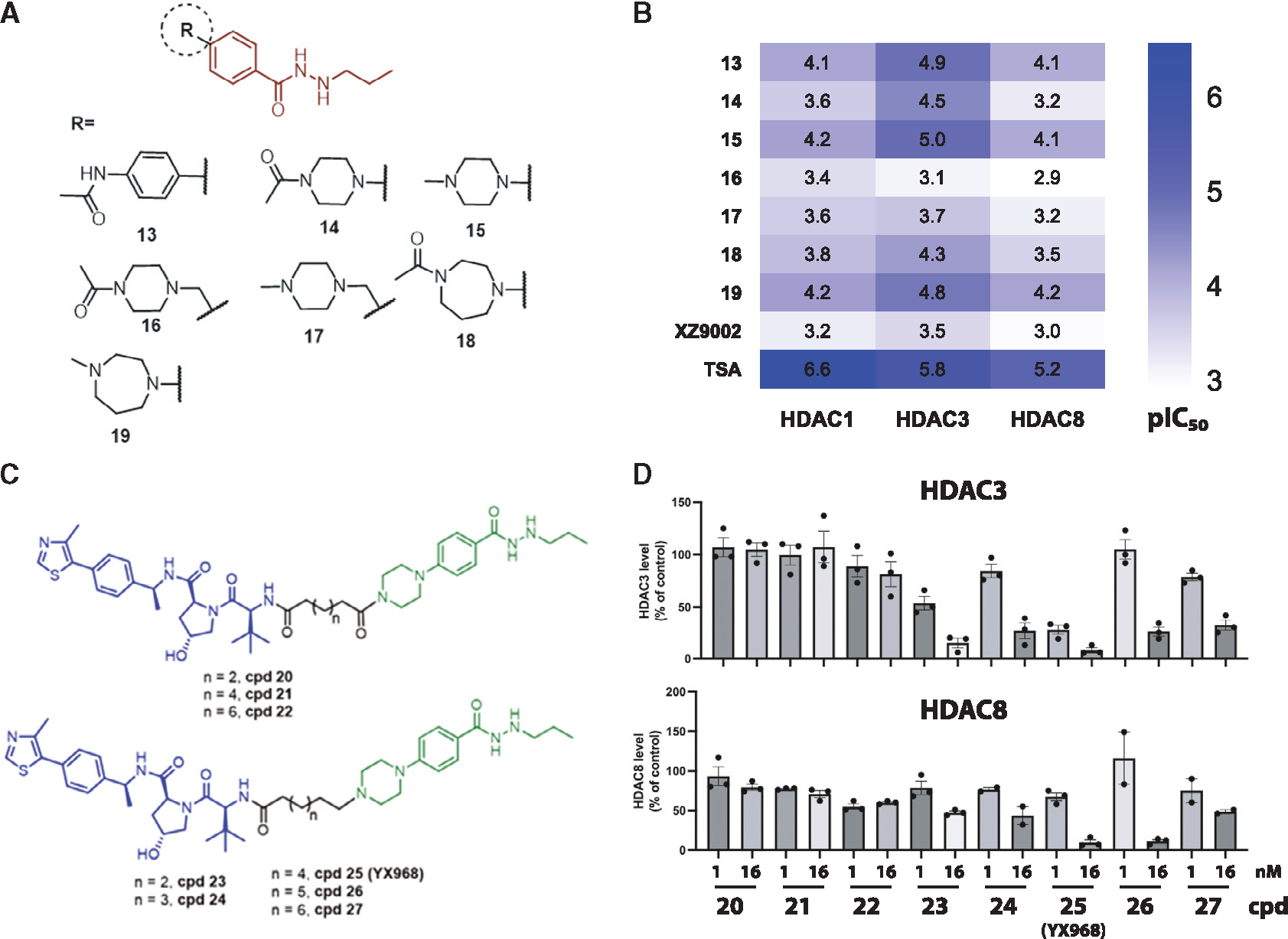
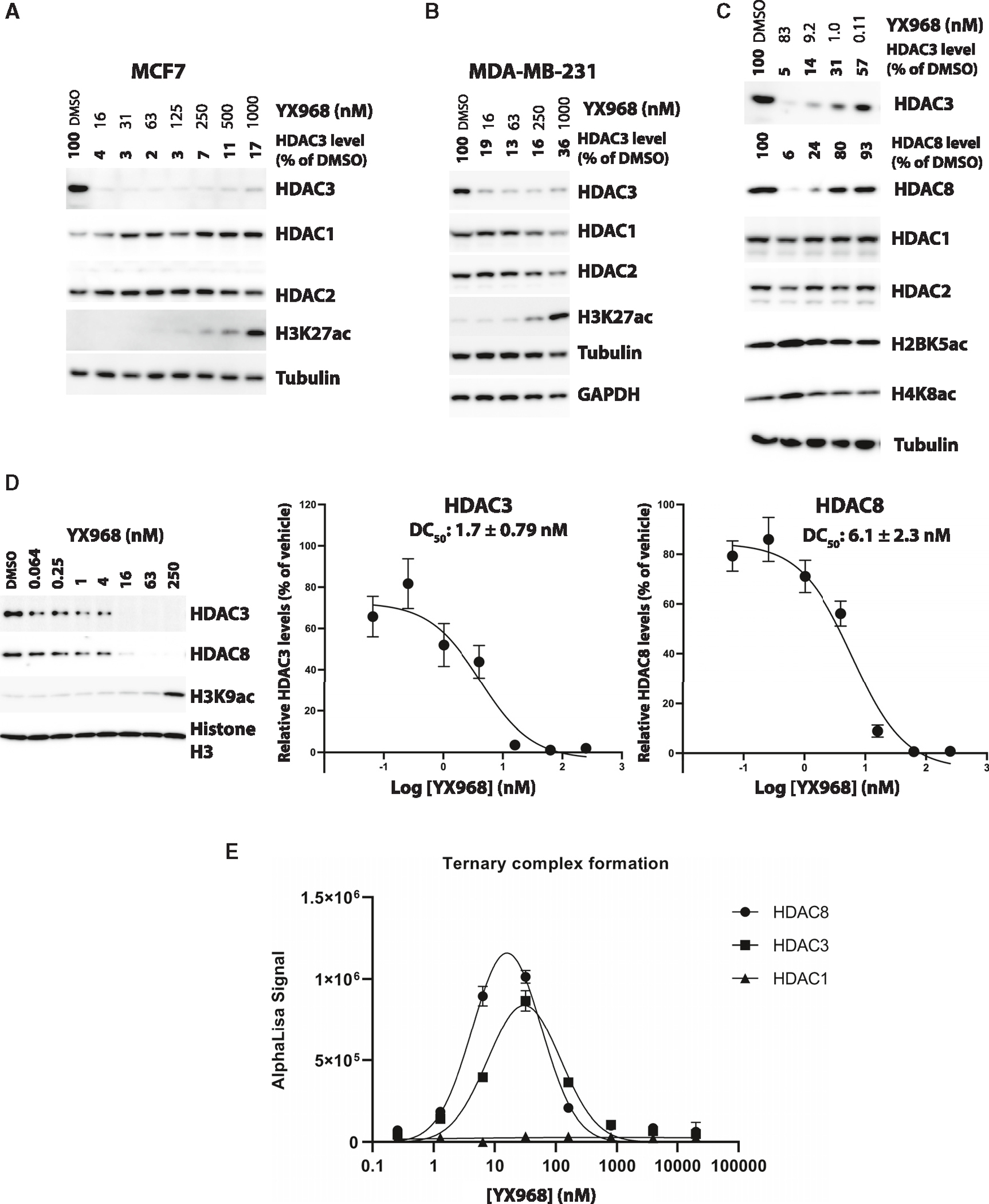
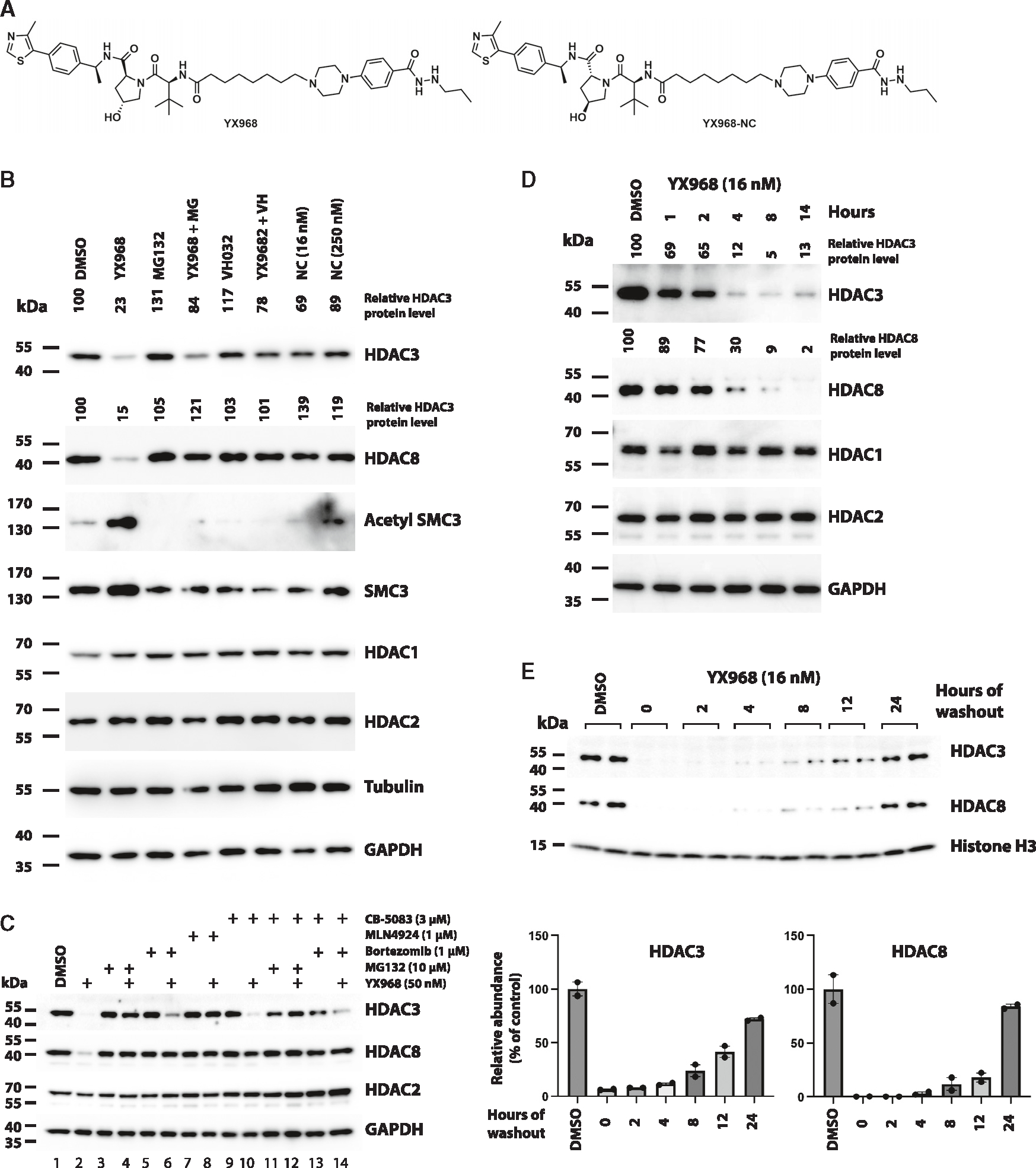
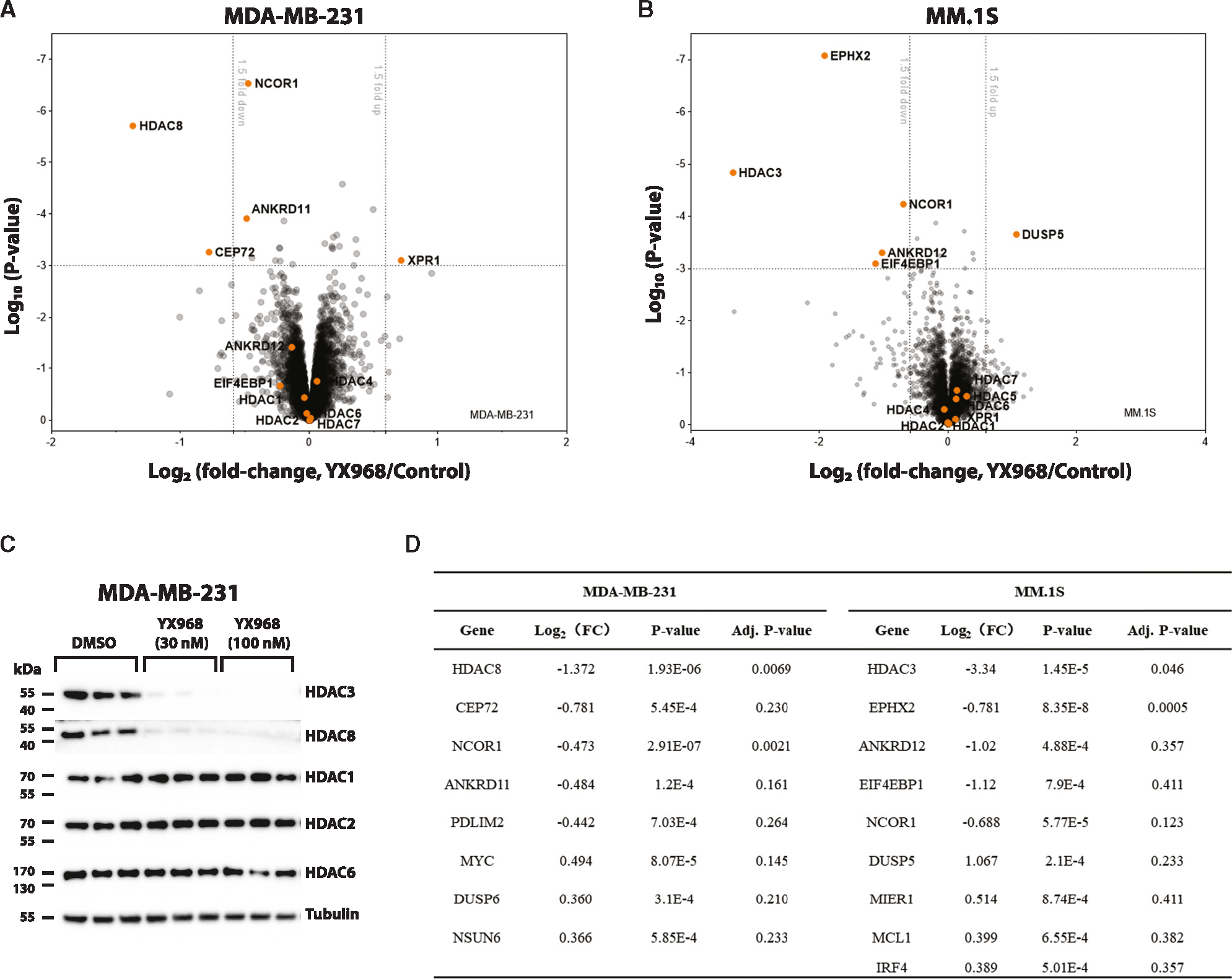
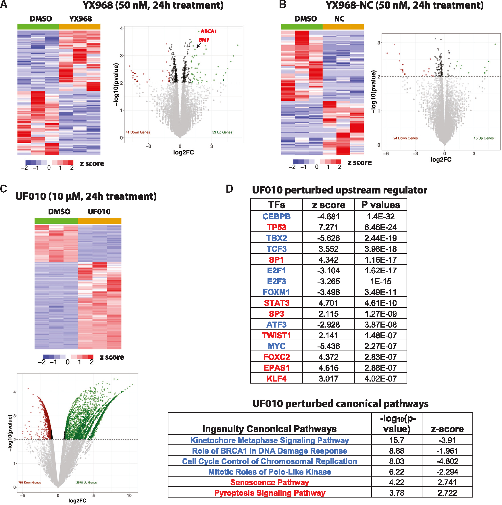
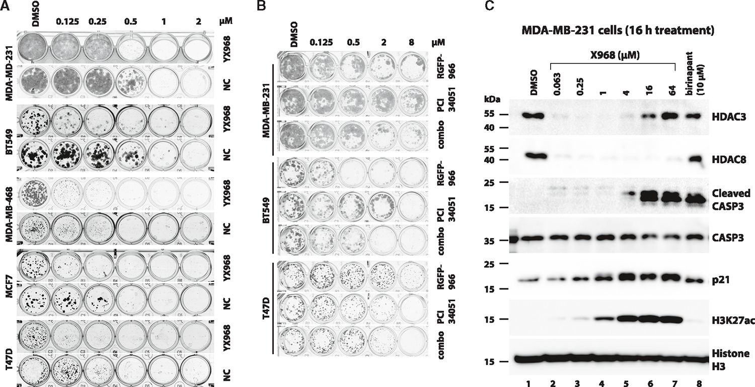
HDAC3 and HDAC8 have critical biological functions and represent highly sought-after therapeutic targets. Because histone deacetylases (HDACs) have a very conserved catalytic domain, developing isozyme-selective inhibitors remains challenging. HDAC3/8 also have deacetylase-independent activity, which cannot be blocked by conventional enzymatic inhibitors. Proteolysis-targeting chimeras (PROTACs) can selectively degrade a target enzyme, abolishing both enzymatic and scaffolding function. Here, we report a novel HDAC3/8 dual degrader YX968 that induces highly potent, rapid, and selective degradation of both HDAC3/8 without triggering pan-HDAC inhibitory effects. Unbiased quantitative proteomic experiments confirmed its high selectivity. HDAC3/8 degradation by YX968 does not induce histone hyperacetylation and broad transcriptomic perturbation. Thus, histone hyperacetylation may be a major factor for altering transcription. YX968 promotes apoptosis and kills cancer cells with a high potency in vitro. YX968 thus represents a new probe for dissecting the complex biological functions of HDAC3/8.


Conflict of interest statement

Declaration of interests X.Y., X.Z., G.Z., and D. Liao are co-inventors of a patent application related to this study filed on behalf of University of Florida Research Foundation. Other authors declare no competing interests.

Figures

Figure 1.
Figure 1.. Inhibitory activity of modified warheads and degradation activity of PROTACs
(A) The structures of modified HDAC inhibitors. (B) The HDAC1/3/8 inhibitory activities of modified warheads. pIC50 was plotted as a heatmap. Each value is the average of two independent assays. (C) The structures of new PROTACs. (D) MDA-MD-231 cells were treated with the PROTACs for 14 h. Shown are average values ±SEM (n = 2 or 3). See also Table S1 and Figures S1–S3.
Figure 2.
Figure 2.. YX968 selectively degrades HDAC3 and HDAC8
(A–C) Western blots showing the levels of the indicated proteins in MCF7 (A), MDA-MD-231 (B and C) cells treated with YX968 for 14 h. HDAC3/8 band intensity was normalized against that of tubulin in each sample. (D) The dose-response curves and DC50 values of HDAC3/8 degradation in MDA-MB-231 cells (treatment for 8 h). Representative Western blot images are shown. (E) Ternary complex formation between the VHL complex consisting of VHL, elongin B and C, and HDAC1, 3, or 8, and YX968 based on in vitro AlphaLISA assay. Data are shown as mean ± SEM (n = 3). See also Figures S3 and S4.
Figure 3.
Figure 3.. Mechanism underlying YX968-induced degradation of HDAC3 and HDAC8
(A) Chemical structure of XY968 and YX968-NC (the negative control of YX968 that does not bind VHL). (B) MDA-MB-231 cells were pretreated with 1 μM MG132 (MG), or 10 μM VHL ligand VH032 for 1 h followed by adding YX968 (16 nM) for 14 h. Cells were also similarly treated with YX968-NC (Shown as NC in Figure 3B). Western blot was done for detecting the indicated proteins. (C) Additional probes for assessing the mechanism by which YX968 degrades HDAC3/8. (D) Time-course assay in MDA-MB-231 cells after treatment with 16 nM YX968. (E) MDA-MB-231 cells were incubated with 16 nM YX968 for 24 h followed by drug washout, and the treated cells were cultured in drug-free medium for the indicated time points. Samples from two independent assays are loaded. The normalized levels of HDAC3/8 protein are plotted. See also Figures S2 and S4.
Figure 4.
Figure 4.. Proteomic profiling of YX968-mediaded degradation
(A) A scatterplot depicting the log2(fold change) of relative protein abundance in MDA-MB-231 cells treated with YX968 (100 nM, 2 h) compared to those treated with DMSO in TMT proteomic profiling. (B) A scatterplot depicting the log2(fold change) of relative protein abundance in MM.1S cells treated with YX968 (50 nM, 3 h) compared to those treated with DMSO in diaPASEF based label-free proteomic profiling. (C) Western blot confirmation of HDAC3/8 degradation of the cell lysates used for TMT profiling in three replicates in MDA-MB-231 cells in (A). (D) Top hits identified by TMT (A) and diaPASEF (B) proteomic profiling. See also Table S2.
Figure 5.
Figure 5.. YX968 does not significantly alter transcriptome
(A–C) A heatmap of RNA-seq data and a volcano plot of DEGs in cells treated with YX968 (A), YX968-NC (B), or UF010 (C) for 24 h. (D) IPA pathways (representative upstream regulators and canonical pathways) activated (red types) or inhibited (blue types) by UF010. See also Figure S5.
Figure 6.
Figure 6.. YX968 is highly potent to suppress cancer cell growth
(A) Colony formation assays using the indicated cell lines that were exposed to DMSO, YX968, or YX968-NC at the indicated concentrations. Colonies were fixed and stained after treatment. Representative results of four replicates. (B) Colony formation assays using the indicated cell lines that were exposed to DMSO, RGFP-966, PCI 34051, or a combination (combo) of RGFP-966 + PCI 34051. Representative results of four replicates. (C) YX968 promotes CASP3 activation. MDA-MB-231 cells were treated with YX968 at the indicated concentrations for 16 h. The cells were also treated with the IAP inhibitor birinapant as a control. The lysates of the treated cells were analyzed by Western blot with antibodies against the indicated proteins. See also Figure S6.
Scheme 1.
Scheme 1.. Synthesis of the compounds 14–19a
aReagents and conditions: (a) K2CO3, DMF, 90°C, 12 h; (b) Hydrazine monohydrate, EtOH, reflux, overnight; (c) Propionaldehyde, THF/MeOH, rt, 30 min, then NaBH4, MeOH, rt, 2 h; (d) NaBH(OAc)3, DCM, rt, overnight.
Scheme 2.
Scheme 2.. Synthesis of compounds 20–27a
aReagents and conditions: (a) 1-Cbz-piperazine, K2CO3, DMF, 90°C, 12 h; (b) Hydrazine monohydrate, EtOH, reflux, overnight; (c) Propionaldehyde, THF/MeOH, rt, 30 min, then NaBH4, MeOH, rt, 2 h; (d) Boc2O, TEA, DCM, rt, overnight; (e) H2, Pd/C (10%), MeOH, 4 h; (f) HATU, DIPEA, DCM, rt, overnight; (g) K2CO3, KI, MeCN, 65°C, overnight; (h) TFA, DCM, rt, 2 h.

Similar articles

Cited by

References

    1. Dawson MA (2017). The cancer epigenome: Concepts, challenges, and therapeutic opportunities. Science 355, 1147–1152. 10.1126/science.aam7304. - DOI - PubMed
    1. Gryder BE, Pomella S, Sayers C, Wu XS, Song Y, Chiarella AM, Bagchi S, Chou HC, Sinniah RS, Walton A, et al. (2019). Histone hyperacetylation disrupts core gene regulatory architecture in rhabdomyosarcoma. Nat. Genet. 51, 1714–1722. 10.1038/s41588-019-0534-4. - DOI - PMC - PubMed
    1. Waddell AR, Huang H, and Liao D (2021). CBP/p300: Critical Co-Activators for Nuclear Steroid Hormone Receptors and Emerging Therapeutic Targets in Prostate and Breast Cancers. Cancers 13, 2872. 10.3390/cancers13122872. - DOI - PMC - PubMed
    1. Chen YJC, Koutelou E, and Dent SYR (2022). Now open: Evolving insights to the roles of lysine acetylation in chromatin organization and function. Mol. Cell 82, 716–727. 10.1016/j.molcel.2021.12.004. - DOI - PMC - PubMed
    1. Yang XJ, and Seto E (2008). The Rpd3/Hda1 family of lysine deacetylases: from bacteria and yeast to mice and men. Nat. Rev. Mol. Cell Biol. 9, 206–218. 10.1038/nrm2346. - DOI - PMC - PubMed

Publication types

MeSH terms