. 2023;16(5):847-856.
doi: 10.1016/j.jcmgh.2023.08.003. Epub 2023 Aug 10.

GLP-2 Improves Hepatic Inflammation and Fibrosis in Mdr2-/- Mice Via Activation of NR4a1/Nur77 in Hepatic Stellate Cells and Intestinal FXR Signaling

Affiliations

GLP-2 Improves Hepatic Inflammation and Fibrosis in Mdr2-/- Mice Via Activation of NR4a1/Nur77 in Hepatic Stellate Cells and Intestinal FXR Signaling

Claudia D Fuchs et al. Cell Mol Gastroenterol Hepatol. 2023.

Abstract

Background & aims: Glucagon-like peptide (GLP)-2 may exert antifibrotic effects on hepatic stellate cells (HSCs). Thus, we aimed to test whether application of the GLP-2 analogue teduglutide has hepatoprotective and antifibrotic effects in the Mdr2/Abcb4-/- mouse model of sclerosing cholangitis displaying hepatic inflammation and fibrosis.

Methods: Mdr2-/- mice were injected daily for 4 weeks with teduglutide followed by gene expression profiling (bulk liver; isolated HSCs) and immunohistochemistry. Activated HSCs (LX2 cells) and immortalized human hepatocytes and human intestinal organoids were treated with GLP-2. mRNA profiling by reverse transcription polymerase chain reaction and electrophoretic mobility shift assay using cytosolic and nuclear protein extracts was performed.

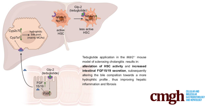
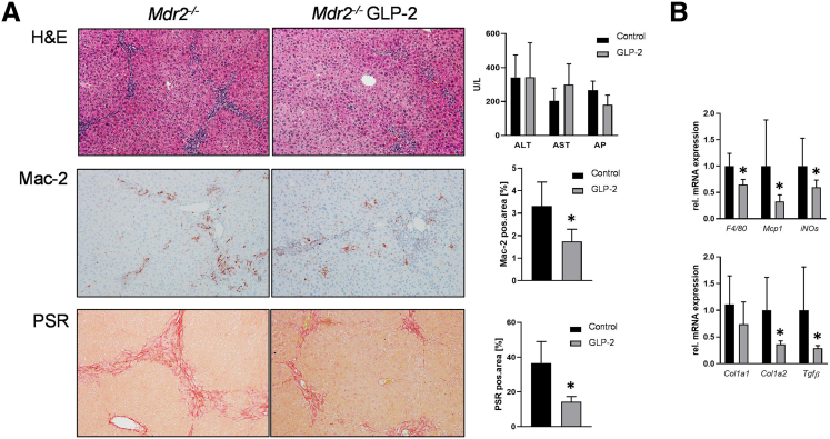
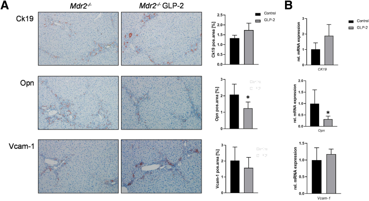
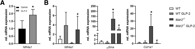
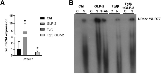
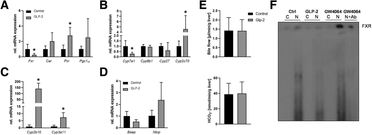
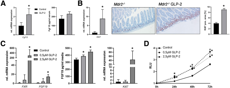
Results: Hepatic inflammation, fibrosis, and reactive cholangiocyte phenotype were improved in GLP-2-treated Mdr2-/- mice. Primary HSCs isolated from Mdr2-/- mice and LX2 cells exposed to GLP-2 in vitro displayed significantly increased mRNA expression levels of NR4a1/Nur77 (P < .05). Electrophoretic mobility shift assay revealed an increased nuclear NR4a1 binding after GLP-2 treatment in LX2 cells. Moreover, GLP-2 alleviated the Tgfβ-mediated reduction of NR4a1 nuclear binding activity. In vivo, GLP-2 treatment of Mdr2-/- mice resulted in increased intrahepatic levels of muricholic acids (accordingly Cyp2c70 mRNA expression was significantly increased), and in reduced mRNA levels of Cyp7a1 and FXR. Serum Fgf15 levels were increased in Mdr2-/- mice treated with GLP-2. Accordingly, GLP-2 treatment of human intestinal organoids activated their FXR-FGF19 signaling axis.

Conclusions: GLP-2 treatment increased NR4a1/Nur77 activation in HSCs, subsequently attenuating their activation. GLP-2 promoted intestinal Fxr-Fgf15/19 signaling resulting in reduced Cyp7a1 and increased Cyp2c70 expression in the liver, contributing to hepatoprotective and antifibrotic effects of GLP-2 in the Mdr2-/- mouse model.

Keywords: Bile Acid Homeostasis; FGF15/19; Fibrosis; Nuclear Binding.


Figures

None
Graphical abstract
Figure 1
Figure 1
GLP-2 treatment improves hepatic inflammation and fibrosis in Mdr2-/-mice. (A) Representative hematoxylin-eosin images (×10 magnification) with markedly improved liver histology in Mdr2-/- mice treated with GLP-2. Serum biochemistry reflects unchanged levels of transaminases (alanine aminotransferase, aspartate aminotransferase) and alkaline phosphatase. Representative Mac-2 and Sirius red (SR) pictures (×10 magnification) show reduced hepatic inflammation and fibrosis in Mdr2-/- mice treated with GLP-2. (B) Real-time polymerase chain reaction was used to assess the mRNA expression of inflammatory and fibrotic markers F4/80, Mcp1, iNOs, Col1a1, Col1a2, and Tgfβ, which were significantly reduced in Mdr2-/- mice treated with GLP-2. mRNA expression values were normalized against 36b4 levels and are shown relative to the expression level in Mdr2-/- control subjects. ∗Significant difference from Mdr2-/- control mice; P < .05. Computational analysis of histologic pictures was done via Image J 1.51j8. ALT, alanine aminotransferase; AP, alkaline phosphatase; AST, aspartate aminotransferase.
Figure 2
Figure 2
GLP-2 treatment improves reactive cholangiocyte phenotype in Mdr2-/-mice. (A) Representative Ck19 images (×10 magnification) show tendentially increased cholangiocyte proliferation in GLP-2-treated Mdr2-/- animals, whereas representative osteopontin (Opn) images (×10 magnification) reflect reduced Opn secretion from cholangiocytes. Vcam-1 images (×10 magnification) remained unchanged among the groups. (B) Real-time polymerase chain reaction was used to assess the mRNA expression of Ck19, Opn, and Vcam-1. Whereas Ck19 expression tended to be increased, Opn levels were reduced and Vcam-1 remained unchanged in Mdr2-/- mice treated with GLP-2. mRNA expression values were normalized against 36b4 levels and are shown relative to the expression level in Mdr2-/- control animals. ∗Significant difference from Mdr2-/- control mice; P < .05. Computational analysis of histologic pictures was done via Image J 1.51j8.
Figure 3
Figure 3
NR4a1/Nur77 mRNA expression is significantly increased in primary hepatic stellate cells because of GLP-2 treatment. (A) Real-time polymerase chain reaction was used to assess the mRNA expression of NR4a1/Nur77 in whole liver homogenate. mRNA expression values were normalized against 36b4 levels and are shown relative to expression level in Mdr2-/- control subjects. ∗Significant difference from Mdr2-/- control mice; P < .05 (B) Real-time polymerase chain reaction was used to assess the mRNA expression of NR4a1/Nur77 αSma and Col1a1 in primary hepatic stellate cells isolated from wild-type (WT) and Mdr2-/- mice with and without GLP-2 treatment. GLP-2 treatment significantly increased expression levels of Nr4a1/Nur77. αSma and Col1a1 mRNA levels were significantly reduced in primary hepatic stellate cells isolated from Mdr2-/- mice treated with GLP-2 compared with untreated Mdr2-/- mice. mRNA expression values were normalized against 36b4 levels and are shown relative to the expression level in WT control animals. #Significant difference from Mdr2-/- control mice; P < .05.
Figure 4
Figure 4
GLP-2 treatment increases NR4a1/Nur77 expression and nuclear binding in human hepatic stellate cells in vitro. (A) Real-time polymerase chain reaction was used to assess the mRNA expression of NR4a1/Nur77 in the human hepatic stellate cell line LX-2. Tgfβ-induced suppression of NR4a1/Nur77 could be counteracted by GLP-2 treatment. mRNA expression values were normalized against 36b4 levels and are shown relative to expression level in untreated control cells. ∗Significant difference from untreated control (Ctrl) cells. #Significant difference from Tgfβ-treated cells. P < .05. (B) Representative electrophoretic mobility shift assay demonstrated that GLP-2 treatment increases the nuclear binding of NR4A1/NUR77, whereas Tgfβ challenge led to a reduction of the NR4A1/NUR77 nuclear binding. Of note, GLP-2 treatment was able to counteract the Tgfβ-related reduction of the NR4A1/NUR77 nuclear binding. Ab, antibody; C, cytoplasmatic protein fraction; N, nuclear protein fraction.
Figure 5
Figure 5
GLP-2 treatment interferes with bile acid homeostasis in Mdr2-/-mice. Real-time polymerase chain reaction was used to assess the mRNA expression of (A) Fxr, Car, Pxr, and Pgc1α, (B) Cyp7a1, Cyp8b1, Cyp27, and Cyp2c70, (C) Cyp2b10 and Cyp3a11, and (D) Bsep and Ntcp in the liver. mRNA levels of Fxr were significantly lowered, whereas Car, Pxr, and Pgc1α were increased. Cyp7a1 was significantly lowered because of GLP-2 treatment in Mdr2-/- mice, whereas Cyp8b1 and Cyp27 remained unchanged and Cyp2c70 was increased. Gene expression of detoxification enzymes Cyp2b10 and Cyp3a11 was significantly increased because of GLP-2. Although Bsep expression levels tended to be lowered, Ntcp showed a tendential increase. mRNA expression values were normalized against 36b4 levels and are shown relative to the expression level in Mdr2-/- control subjects. ∗Significant difference from Mdr2-/- control mice; P < .05. (E) Hepatobiliary bile flow and bicarbonate (HCO3-) output were assessed. Neither bile flow nor HCO3- output was changed because of GLP-2 treatment. (F) Representative electrophoretic mobility shift assay demonstrated that GLP-2 treatment suppresses the nuclear binding of FXR, whereas GW4064 (FXR agonist) challenge led to an increase of FXR nuclear binding. Ab, antibody; C, cytoplasmatic protein fraction; N, nuclear protein fraction.
Figure 6
Figure 6
GLP-2 treatment activates intestinal FGF15/19 expression. Real-time polymerase chain reaction was used to assess the intestinal mRNA expression of (A) Fgf15 and (B) Ki67. Fgf15 enzyme-linked immunosorbent assay (ELISA) was used to investigate Fgf15 levels in systemic blood of Mdr2-/- mice on GLP-2 treatment (A). Intestinal Fgf15 mRNA and protein expression as well as Ki67 mRNA levels were elevated in Mdr2-/- mice on GLP-2 treatment. Accordingly, Ki67 immunohistochemistry (×10 magnification) (B) showed an increase in Ki67-positive cells numbers in the intestine of Mdr2-/- mice treated with GLP-2. mRNA expression values were normalized against 18sRNA levels and are shown relative to the expression level in Mdr2-/- control animals. ∗Significant difference from Mdr2-/- control mice; P < .05. (C) Real-time polymerase chain reaction was used to assess the mRNA expression of FXR, FGF19, and KI67 in human-derived intestinal organoids treated with GLP-2. FGF19 ELISA was used to assess FGF19 protein concentration in cell culture supernatant. Gene expression of the aforementioned genes was significantly increased by GLP-2 treatment. mRNA expression values were normalized against 18sRNA levels and are shown relative to expression level in untreated control subjects. FGF19 levels were significantly increased in supernatant of organoids treated with 0.5 μM and 2.5 μM GLP-2. ∗Significant difference from untreated control cells; P < .05. (D) CellTiter-Glo Luminescent Cell Viability Assay was performed to assess cell proliferation in human-derived intestinal organoids. GLP-2 significantly increased cell proliferation independent of the used concentrations. ∗Significant difference from untreated control cells; P < .05.

References

    1. Fickert P., Fuchsbichler A., Wagner M., et al. Regurgitation of bile acids from leaky bile ducts causes sclerosing cholangitis in Mdr2 (Abcb4) knockout mice. Gastroenterology. 2004;127:261–274. - PubMed
    1. Fickert P., Pollheimer M.J., Beuers U., et al. Characterization of animal models for primary sclerosing cholangitis (PSC) J Hepatol. 2014;60:1290–1303. - PMC - PubMed
    1. Lazaridis K.N., Strazzabosco M., Larusso N.F. The cholangiopathies: disorders of biliary epithelia. Gastroenterology. 2004;127:1565–1577. - PubMed
    1. Nishio T., Hu R., Koyama Y., et al. Activated hepatic stellate cells and portal fibroblasts contribute to cholestatic liver fibrosis in MDR2 knockout mice. J Hepatol. 2019;71:573–585. - PubMed
    1. Mederacke I., Hsu C.C., Troeger J.S., et al. Fate tracing reveals hepatic stellate cells as dominant contributors to liver fibrosis independent of its aetiology. Nat Commun. 2013;4:2823. - PMC - PubMed

Publication types