

Prevalence and predictors of radiological left common iliac vein compression in asymptomatic patients
- PMID: 37572778
- PMCID: PMC11523471
- DOI: 10.1016/j.jvsv.2023.07.011
Prevalence and predictors of radiological left common iliac vein compression in asymptomatic patients
Abstract
Objective: The aim of this study was to investigate the prevalence of radiological left common iliac vein (LCIV) compression among the asymptomatic population and identify possible predictors.
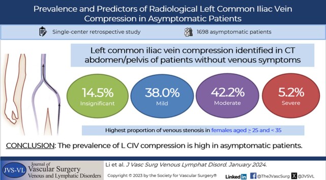
Methods: Contrast-enhanced abdominal and/or pelvic computed tomography scans of eligible asymptomatic patients were examined. The LCIV diameter was measured from different horizontal planes in the venous phase using PACSView. Degree of LCIV compression (Dc) was calculated by a predefined formula and graded as insignificant (Dc < 25%), mild (≥25% Dc < 50%), moderate (≥50% Dc <75%), and severe (Dc ≥ 75%). Venous stenosis was defined as a Dc of ≥50%. Comparison of variables, including gender, age, body mass index (BMI), and comorbidities was performed between the different grades of LCIV compression.
Results: Between November 2019 and July 2022, 1698 eligible asymptomatic patients (53.1% females; mean age, 39.3 ± 11.8 years; mean BMI, 22.9 ± 3.6 kg/m2) were reviewed. The mean Dc was 46.2% (range, 0.29%-90.4%). Insignificant, mild, moderate, and severe compression were distributed in 14.5%, 38.0%, 42.2%, and 5.2% of the cohort population, respectively. Prevalence of venous stenosis was higher in females than males (58.1% vs 42.2%; χ2 = 15.52; P < .001). Females aged ≥25 and <35 years accounted for the highest proportion of venous stenosis than other age groups and was a significant predictor (odds ratio [OR], 3.18; 95% confidence interval [CI], 1.74-7.79; P < .001). In the Asian BMI classification group, being underweight is associated with venous stenosis (OR, 4.69; 95% CI, 2.70-8.14; P < .001) and obesity may be a protective factor (OR, 0.38; 95% CI, 0.23-0.64; P < .001). There is an inverse relationship between Dc and age and BMI.
Conclusions: The prevalence of radiological LCIV compression on computed tomography scans was high, but all patients were asymptomatic. Female gender, especially those aged ≥25 and <35 years, and underweight were possible predictors for venous stenosis.
Keywords: Chronic venous insufficiency; Left common iliac vein compression; May-Thurner syndrome; Radiology; Venous thromboembolism.
Copyright © 2023 The Authors. Published by Elsevier Inc. All rights reserved.
Figures


References
-
- McMurrich J.P. The occurrence of congenital adhesions in the common iliac veins and their relation to thrombosis of the femoral and iliac veins. Am J Med Sci. 1908;135:342–346.
-
- Ehrich W.E., Krumbhaar E.B. A frequent obstructive anomaly of the mouth of the left common iliac vein. Am Heart J. 1943;26:737–750.
-
- MAY R., THURNER J. The cause of the predominantly sinistral occurrence of thrombosis of the pelvic veins. Angiology. 1957;8:419–427. - PubMed
Publication types
MeSH terms
LinkOut - more resources
Full Text Sources
Medical
