

Mini-Review: Enteric glia of the tumor microenvironment: An affair of corruption
- PMID: 37572875
- PMCID: PMC10967235
- DOI: 10.1016/j.neulet.2023.137416
Mini-Review: Enteric glia of the tumor microenvironment: An affair of corruption
Abstract
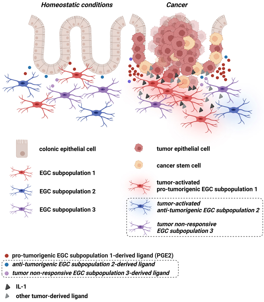
The tumor microenvironment corresponds to a complex mixture of bioactive products released by local and recruited cells whose normal functions have been "corrupted" by cues originating from the tumor, mostly to favor cancer growth, dissemination and resistance to therapies. While the immune and the mesenchymal cellular components of the tumor microenvironment in colon cancer have been under intense scrutiny over the last two decades, the influence of the resident neural cells of the gut on colon carcinogenesis has only very recently begun to draw attention. The vast majority of the resident neural cells of the gastrointestinal tract belong to the enteric nervous system and correspond to enteric neurons and enteric glial cells, both of which have been understudied in the context of colon cancer development and progression. In this review, we especially discuss available evidence on enteric glia impact on colon carcinogenesis. To highlight "corrupted" functioning in enteric glial cells of the tumor microenvironment and its repercussion on tumorigenesis, we first review the main regulatory effects of enteric glial cells on the intestinal epithelium in homeostatic conditions and we next present current knowledge on enteric glia influence on colon tumorigenesis. We particularly examine how enteric glial cell heterogeneity and plasticity require further appreciation to better understand the distinct regulatory interactions enteric glial cell subtypes engage with the various cell types of the tumor, and to identify novel biological targets to block enteric glia pro-carcinogenic signaling.
Keywords: Cancer stem cell; Colon cancer; Enteric glial cell; Intestinal stem cell; Tumor microenvironment.
Copyright © 2023 Elsevier B.V. All rights reserved.
Conflict of interest statement
Declaration of Competing Interest The authors declare that they have no known competing financial interests or personal relationships that could have appeared to influence the work reported in this paper.
Figures
Similar articles
-
Functional Intraregional and Interregional Heterogeneity between Myenteric Glial Cells of the Colon and Duodenum in Mice.J Neurosci. 2022 Nov 16;42(46):8694-8708. doi: 10.1523/JNEUROSCI.2379-20.2022. Epub 2022 Nov 1. J Neurosci. 2022. PMID: 36319118 Free PMC article.
-
Enteric glia: Diversity or plasticity?Brain Res. 2018 Aug 15;1693(Pt B):140-145. doi: 10.1016/j.brainres.2018.02.001. Epub 2018 Feb 7. Brain Res. 2018. PMID: 29425908 Review.
-
Tumor cells hijack enteric glia to activate colon cancer stem cells and stimulate tumorigenesis.EBioMedicine. 2019 Nov;49:172-188. doi: 10.1016/j.ebiom.2019.09.045. Epub 2019 Oct 26. EBioMedicine. 2019. PMID: 31662289 Free PMC article.
-
The Physiology of Enteric Glia.Annu Rev Physiol. 2025 Feb;87(1):353-380. doi: 10.1146/annurev-physiol-022724-105016. Epub 2025 Feb 3. Annu Rev Physiol. 2025. PMID: 39546562 Review.
-
Mini-review: Enteric glial cell heterogeneity: Is it all about the niche?Neurosci Lett. 2023 Aug 24;812:137396. doi: 10.1016/j.neulet.2023.137396. Epub 2023 Jul 12. Neurosci Lett. 2023. PMID: 37442521 Review.
Cited by
-
From diversity to disease: unravelling the role of enteric glial cells.Front Immunol. 2024 Jun 18;15:1408744. doi: 10.3389/fimmu.2024.1408744. eCollection 2024. Front Immunol. 2024. PMID: 38957473 Free PMC article. Review.
-
Exploring the Role of Cellular Interactions in the Colorectal Cancer Microenvironment.J Immunol Res. 2025 Apr 11;2025:4109934. doi: 10.1155/jimr/4109934. eCollection 2025. J Immunol Res. 2025. PMID: 40255905 Free PMC article. Review.
References
Publication types
MeSH terms
Grants and funding
LinkOut - more resources
Full Text Sources
Molecular Biology Databases
