

A novel long non-coding RNA XLOC_004787, is associated with migration and promotes cancer cell proliferation by downregulating mir-203a-3p in gastric cancer
- PMID: 37573302
- PMCID: PMC10422700
- DOI: 10.1186/s12876-023-02912-2
A novel long non-coding RNA XLOC_004787, is associated with migration and promotes cancer cell proliferation by downregulating mir-203a-3p in gastric cancer
Abstract
Background: Long noncoding RNAs (lncRNAs) have been identified as important regulatory factors implicated in a wide array of diseases, including various forms of cancer. However, the roles of most lncRNAs in the progression of gastric cancer (GC) remain largely unexplored. This study investigates the biological function and underlying mechanism of a novel lncRNA, XLOC_004787 in GC.
Methods: The location of XLOC_004787 in GES-1 cells and HGC-27 cells were detected by fluorescence in situ hybridization (FISH) assay. The expression levels of XLOC_004787 were assessed using quantitative real-time fluorescence PCR (qRT-PCR) in various cell lines, including GES-1, MGC-803, MKN-45, BGC-823, SGC-7901, and HGC-27 cells. Functional assays such as Transwell migration, cell counting kit-8 (CCK-8), and colony formation experiments were employed to analyze the effects of XLOC_004787 and miR-203a-3p on cell migration and proliferation. Protein levels associated with GC in these cell lines were examined by Western blotting. The intracellular localization of β-catenin and P-Smad2/3 was assessed using immunofluorescence (IF) assay. Additionally, the interaction between XLOC_004787 and miR-203a-3p was investigated using a dual luciferase assay.
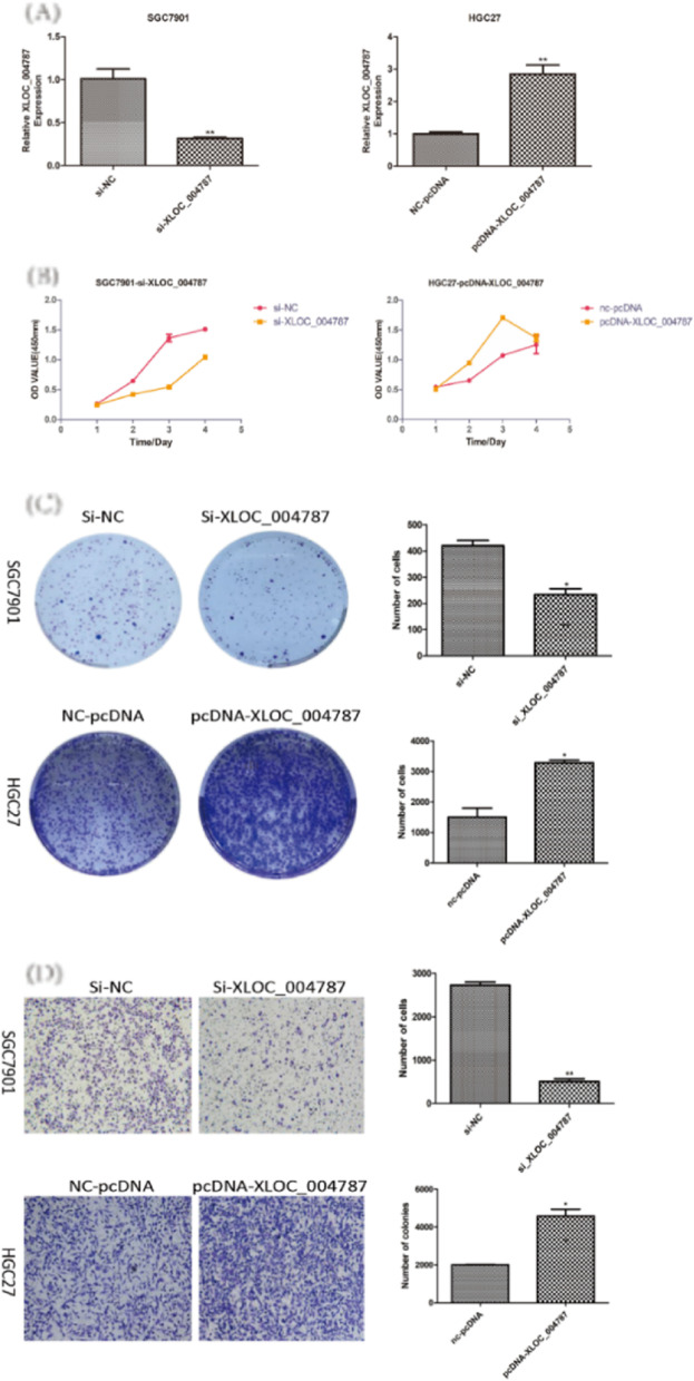
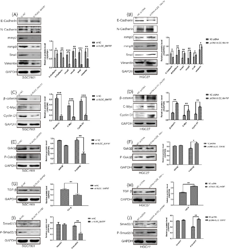
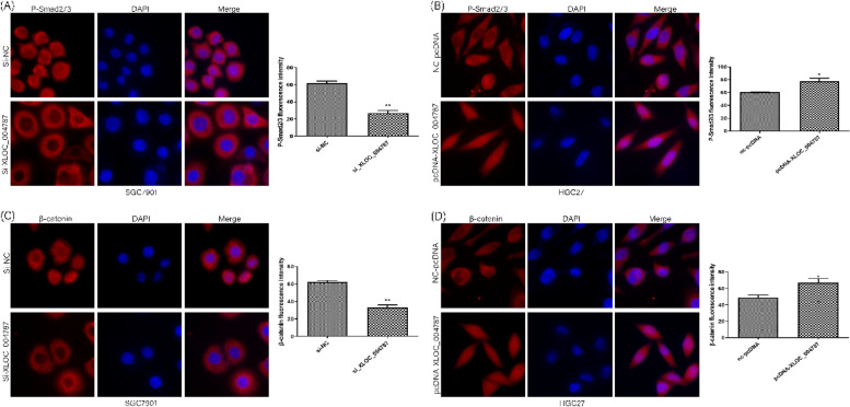
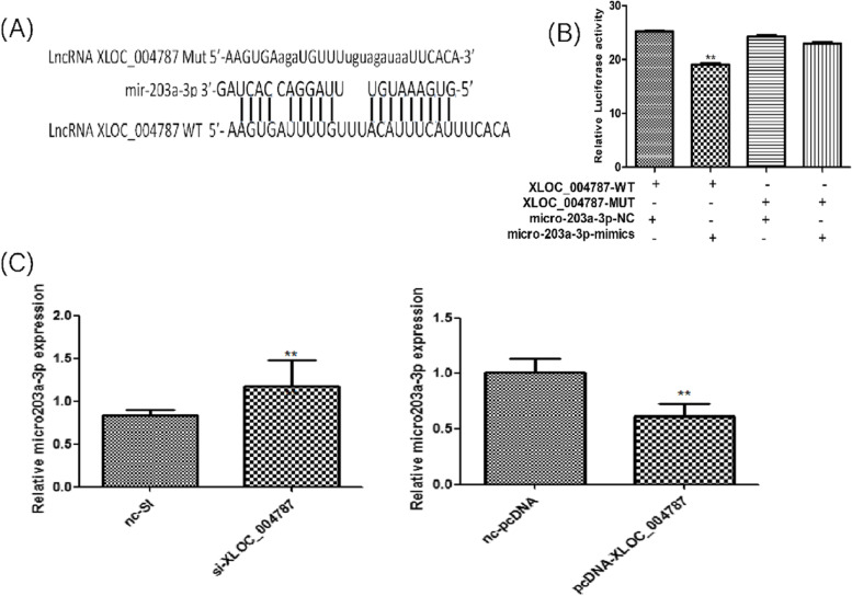
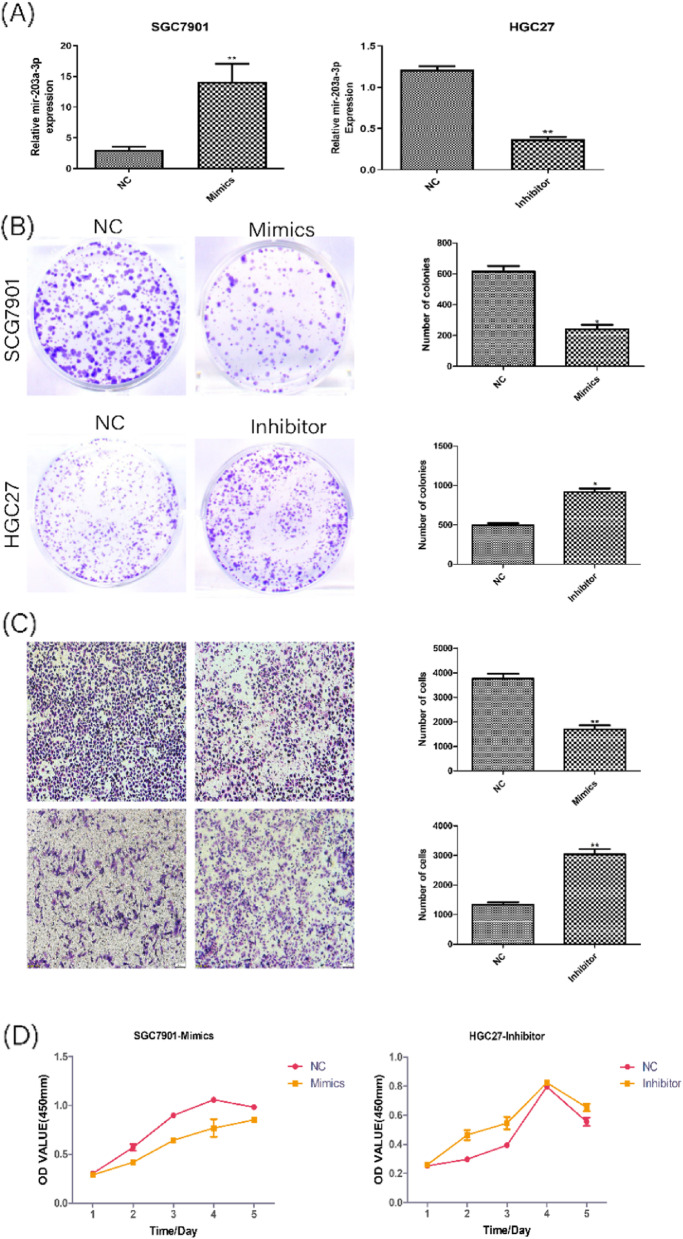
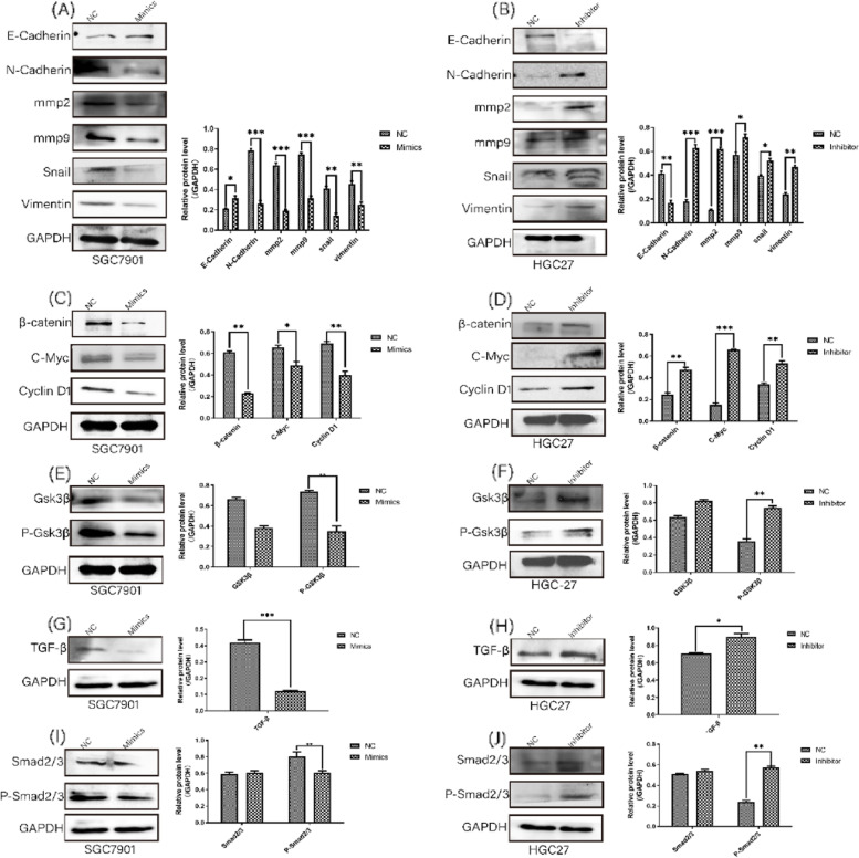
Results: XLOC_004787 was localized at both the cytoplasm and nucleus of GES-1 cells and HGC-27 cells. Compared to normal tissues and GES-1 cells, XLOC_004787 expression was significantly upregulated in GC tissues and cells, with the highest and lowest expression observed in SGC-7901 and HGC-27 cells, respectively. Furthermore, a reduced expression of XLOC_004787 was seen to inhibit migration and proliferation in SGC-7901 cells. Western blotting analysis revealed that a decrease in XLOC_004787 expression correspondingly decreased the expression of N-cadherin, mmp2, mmp9, Snail, Vimentin, β-catenin, C-myc, Cyclin D1, and TGF-β, while concurrently increasing E-cadherin expression. This was also associated with diminished expression of P-Smad2/3 in relation to Smad2/3, and reduced P-Gsk3β expression in comparison to Gsk3β. Additionally, the nuclear entry of P-Smad2/3 and β-catenin was reduced by lower XLOC_004787 expression. Amplifying XLOC_004787 expression via pcDNA_XLOC_004787 suggested a potential for cancer promotion. Notably, XLOC_004787 was found to negatively regulate mir-203a-3p expression, with potential binding sites identified between the two. Higher mir-203a-3p expression was observed to decrease migration and proliferation, and enhance E-cadherin expression. Conversely, suppression of mir-203a-3p expression suggested a potential promotion of proliferation and migration in GC cells.
Conclusions: These results suggest that XLOC_004787, found to be upregulated in GC tissues, potentially promotes proliferation and migration in GC cells. This occurs through the activation of TGF-β and Wnt/β-catenin signaling pathways and the expression of EMT-related proteins. Additionally, XLOC_004787 may influence cell migration and proliferation by modulating the signaling pathway via the adsorption and inhibition of mir-203a-3p.
Keywords: Gastric cancer; Migration; Mir-203a-3p; Proliferation; XLOC_004787.
© 2023. BioMed Central Ltd., part of Springer Nature.
Conflict of interest statement
There is no conflict of interest in the publication of this article.
Figures

Similar articles
-
MiR-203a-3p regulates the biological behaviors of ovarian cancer cells through mediating the Akt/GSK-3β/Snail signaling pathway by targeting ATM.J Ovarian Res. 2019 Jul 5;12(1):60. doi: 10.1186/s13048-019-0532-2. J Ovarian Res. 2019. PMID: 31277702 Free PMC article.
-
LncRNA HOTAIR is a Prognostic Biomarker for the Proliferation and Chemoresistance of Colorectal Cancer via MiR-203a-3p-Mediated Wnt/ß-Catenin Signaling Pathway.Cell Physiol Biochem. 2018;46(3):1275-1285. doi: 10.1159/000489110. Epub 2018 Apr 16. Cell Physiol Biochem. 2018. PMID: 29680837
-
Tumor Inhibitory Effect of Long Non-coding RNA LOC100505817 on Gastric Cancer.Pathol Oncol Res. 2021 May 26;27:581542. doi: 10.3389/pore.2021.581542. eCollection 2021. Pathol Oncol Res. 2021. PMID: 34385891 Free PMC article.
-
MicroRNA-205-5p inhibits the growth and migration of breast cancer through targeting Wnt/β-catenin co-receptor LRP6 and interacting with lncRNAs.Mol Cell Biochem. 2025 Apr;480(4):2117-2129. doi: 10.1007/s11010-024-05136-4. Epub 2024 Oct 26. Mol Cell Biochem. 2025. PMID: 39461917 Review.
-
miR-203a-A multifaceted regulator modulating cancer hallmarks and therapy response.IUBMB Life. 2024 Mar;76(3):108-124. doi: 10.1002/iub.2786. Epub 2023 Oct 4. IUBMB Life. 2024. PMID: 37792370 Review.
Cited by
-
The Role of the Transforming Growth Factor-β Signaling Pathway in Gastrointestinal Cancers.Biomolecules. 2023 Oct 19;13(10):1551. doi: 10.3390/biom13101551. Biomolecules. 2023. PMID: 37892233 Free PMC article. Review.
-
Revealing the Potential of Solamargine for Anti Metastasis and Angiogenesis Inhibition in Nasopharyngeal Carcinoma.J Inflamm Res. 2025 Apr 8;18:4879-4898. doi: 10.2147/JIR.S485244. eCollection 2025. J Inflamm Res. 2025. PMID: 40224395 Free PMC article.
-
MiR-424-5p suppresses tumor growth and progression by directly targeting CHEK1 and activating cell cycle pathway in Hepatocellular Carcinoma.Heliyon. 2024 Sep 11;10(18):e37769. doi: 10.1016/j.heliyon.2024.e37769. eCollection 2024 Sep 30. Heliyon. 2024. PMID: 39309825 Free PMC article.
-
A novel tRNA-derived fragment tRF-17-18VBY9M works as a potential diagnostic biomarker for gastric cancer.J Cancer Res Clin Oncol. 2024 May 20;150(5):263. doi: 10.1007/s00432-024-05792-5. J Cancer Res Clin Oncol. 2024. PMID: 38767702 Free PMC article.
-
A bioinformatics analysis and experimental validation of PDGFD as a promising diagnostic biomarker for acute myeloid leukemia.Sci Rep. 2025 Apr 28;15(1):14862. doi: 10.1038/s41598-025-99038-0. Sci Rep. 2025. PMID: 40295666 Free PMC article.
References
MeSH terms
Substances
LinkOut - more resources
Full Text Sources
Medical
Research Materials
Miscellaneous
