Review
. 2023 Aug 12;23(1):563.
doi: 10.1186/s12903-023-03259-6.

Metastasising ameloblastoma or ameloblastic carcinoma? A case report with mutation analyses

Affiliations
Review

Metastasising ameloblastoma or ameloblastic carcinoma? A case report with mutation analyses

Pavel Hurník et al. BMC Oral Health. .

Abstract

Background: Ameloblastic carcinoma and metastasising ameloblastoma are rare epithelial odontogenic tumours with aggressive features. Distinguishing between these two lesions is often clinically difficult but necessary to predict tumour behaviour or to plan future therapy. Here, we provide a brief review of the literature available on these two types of lesions and present a new case report of a young man with an ameloblastoma displaying metastatic features. We also use this case to illustrate the similarities and differences between these two types of tumours and the difficulties of their differential diagnosis.

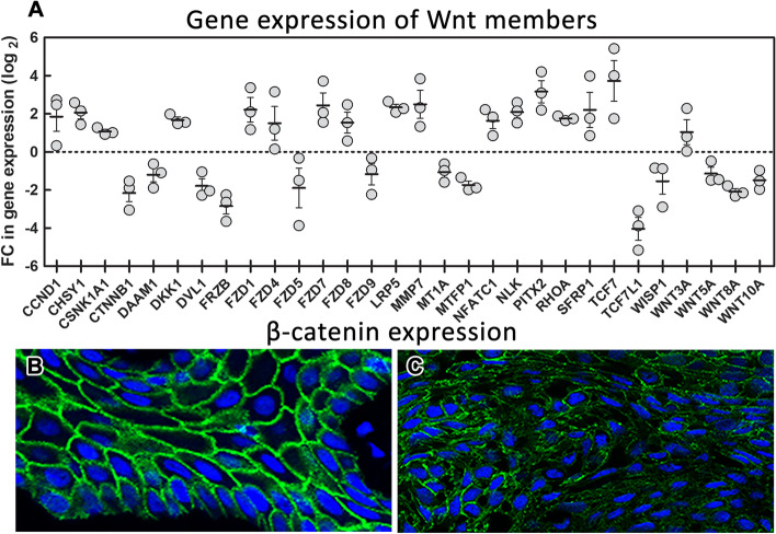
Case presentation: Our histopathological analyses uncovered a metastasising tumour with features of ameloblastic carcinoma, which developed from the ameloblastoma. We profiled the gene expression of Wnt pathway members in ameloblastoma sample of this patient, because multiple molecules of this pathway are involved in the establishing of cell polarity, cell migration or for epithelial-mesenchymal transition during tumour metastasis to evaluate features of tumor behaviour. Indeed, we found upregulation of several cell migration-related genes in our patient. Moreover, we uncovered somatic mutation BRAF p.V600E with known pathological role in cancerogenesis and germline heterozygous FANCA p.S858R mutation, whose interpretation in this context has not been discussed yet.

Conclusions: In conclusion, we have uncovered a unique case of ameloblastic carcinoma associated with an alteration of Wnt signalling and the presence of BRAF mutation. Development of harmful state of our patient might be also supported by the germline mutation in one FANCA allele, however this has to be confirmed by further analyses.

Keywords: Ameloblastoma; BRAF; Benign; Carcinoma; Case report; FANCA; Malignant; Metastasizing; Wnt pathway.


Conflict of interest statement

The authors declare that the research was conducted in the absence of any commercial or financial relationships that could be construed as a potential conflict of interest.

The authors declare that they have no known competing financial interests or personal relationships that could have appeared to influence the work reported in this paper.

Figures

Fig. 1
Fig. 1
Timeline of diagnosis. Patient’s timeline with the important diagnostic events, treatment and collection of samples for sequencing between years 2012 to 2022
Fig. 2
Fig. 2
RTG and CT analysis and immunohistochemistry of the first collected oral sample of our patient. First X-ray of patient with ameloblastoma, where is retained wisdom tooth and loss of bone at the right side of mandible (A). 3D reconstruction of mandible from CT scans. There is a large lesion with bone resorption primary in the right side of the mandible extending into the region of the mandibular ramus (B). Epithelial cords of tumourous components with columnar cells exhibiting reverse polarity; Haematoxylin–Eosin staining, magnification 200x (C). Diffuse membranous positivity of pan-cytokeratin in tumourous cells; immunohistochemistry: CK AE1/AE3 (pan-cytokeratin (1:1, cat. no. 961, Abcam, UK), magnification 200x (D). Minimal nuclear immunoreactivity of p53 in tumour cells; immunohistochemistry: p53, magnification 200x (E). Slight nuclear positivity in Ki67 staining displays a weak proliferation activity of tumorous tissue; immunohistochemistry: Ki67 (1:200, cat. no. 275-R-16, Cell Marque, USA), magnification 200x (F). 3,3′-diaminobenzidine (DAB, cat. No. K3468, DAKO, Agilent Technologies, USA) was used to detect positive cells and Haematoxylin was applied to counterstain the nuclei
Fig. 3
Fig. 3
MRI and immunohistochemistry of the oral tissue sample after the tumor recurrence. MRI of head tumour recurrence into the region of the right zygomatic bone growing into the caudal part of the orbit (A). Epithelial cords of tumour compound of columnar cells with reverse polarization, dense cellularity in basal parts of tumour, increased mitotic activity and nuclear hyperchromasia; Haematoxylin–Eosin staining, magnification 200x (B). Diffuse membranous positivity of pan-cytokeratin with maximum of positivity in stellate reticulum-like cells in tumour tissue; immunohistochemistry: CK AE1/AE3 (pan-cytokeratin (1:1, cat. no. 961, Abcam, UK), magnification 200x (C). Weak nuclear immunoreactivity of p53 in tumour cell; immunohistochemistry: p53, magnification 200x (D). Nuclear positivity of Ki67 staining reaches the level up to 10% and represents increased proliferation activity; immunohistochemistry: Ki67 (1:200, cat. no. 275-R-16, Cell Marque, USA), magnification 200x (E). 3,3′-diaminobenzidine (DAB, cat. No. K3468, DAKO, Agilent Technologies, USA) was used to detect the positive cells and Haematoxylin was applied to counterstain the nuclei
Fig. 4
Fig. 4
CT and PET CT analysis and immunohistochemistry of the lung metastases. CT of lungs with suspicious signs of tumour in the S3 region of the left lung (A). PET CT confirmed viable tumorous tissue in the S3 region of left lung (B). Epithelial cords located in dense fibrovascular stroma in lung tissue with features including high N/C ratio, increased mitotic figures. These features are suspicious for possible malignant behaviour of tumour, but not fully represent malignancy; Haematoxylin–Eosin staining, magnification 200x (C). Diffuse membranous positivity of pan-cytokeratin with the maximum of positivity in stellate reticulum-like cells of tumour. Immunohistochemistry—CK AE1/AE3 (pan-cytokeratin (1:1, cat. no. 961, Abcam, UK), magnification 200x (D). Nuclear staining of p53 in hot-spots; immunohistochemistry p53, magnification 200x (E). Nuclear positivity of Ki67 staining is increased up to 15–20% in cell-dense hot-spots and represents increased proliferation activity of the tissue; immunohistochemistry Ki67 (1:200, cat. no. 275-R-16, Cell Marque, USA), magnification 200x (F). 3,3′-diaminobenzidine (DAB, cat. No. K3468, DAKO, Agilent Technologies, USA) was used to detect the positive cells and Haematoxylin was applied to counterstain the nuclei
Fig. 5
Fig. 5
RNA expression analysis of Wnt related genes and β-catenin immunofluorescent detection. Expression analysis of genes related to Wnt signalling pathway. Ameloblastoma tumour tissue sample was compared to three healthy gingival tissues (represented by three data points for each gene). Values for gene expression changes were calculated using 2-ΔΔCT method and subsequently log2 transformed (A). In the control oral epithelial cells, β-catenin signal is membranous, and it is regularly distributed through cell surfaces (B). In ameloblastic carcinoma, there is a signal loss in the tumorous cells and the expression pattern is discontinuous and stippled (C). Immunofluorescence: β-catenin (6B3) (beta-catenin (1:100, cat. no. 9582, Cell Signalling Technology, USA), the nuclei were counterstained by DRAQ5™ (cat. no. 62251, Thermo Fisher Scientific)

References

    1. Wright JM, Vered M. Update from the 4th Edition of the world health organization classification of head and neck tumours: odontogenic and maxillofacial bone Tumors. Head Neck Pathol. 2017;11(1):68–77. - PMC - PubMed
    1. Kramer IR, Pindborg JJ, Shear M. The WHO Histological Typing of Odontogenic Tumours. A commentary on the Second Edition. Cancer. 1992;70(12):2988–94. - PubMed
    1. Kishore M, Panat SR, Aggarwal A, Upadhyay N, Agarwal N. Ameloblastic Carcinoma: A Case Report. J Clin Diagn Res. 2015;9(7):ZD27–8. - PMC - PubMed
    1. Palanisamy JC, Jenzer AC. Ameloblastoma. StatPearls. Treasure Island (FL)2022.
    1. Jing W, Xuan M, Lin Y, Wu L, Liu L, Zheng X, et al. Odontogenic tumours: a retrospective study of 1642 cases in a Chinese population. Int J Oral Maxillofac Surg. 2007;36(1):20–25. - PubMed

Substances