. 2023 Aug 12;16(1):94.
doi: 10.1186/s13045-023-01487-5.

Synergistic efficacy of simultaneous anti-TGF-β/VEGF bispecific antibody and PD-1 blockade in cancer therapy

Affiliations

Synergistic efficacy of simultaneous anti-TGF-β/VEGF bispecific antibody and PD-1 blockade in cancer therapy

Mengke Niu et al. J Hematol Oncol. .

Abstract

Background: Recently, therapeutic antibodies against programmed cell death 1 (PD-1) and its ligand (PD-L1) have exerted potent anticancer effect in a variety of tumors. However, blocking the PD-1/PD-L1 axis alone is not sufficient to restore normal immune response. Other negative regulators of antitumor immunity, like TGF-β and VEGFA, are also involved in immune escape of tumor cells and induce immunotherapy resistance.

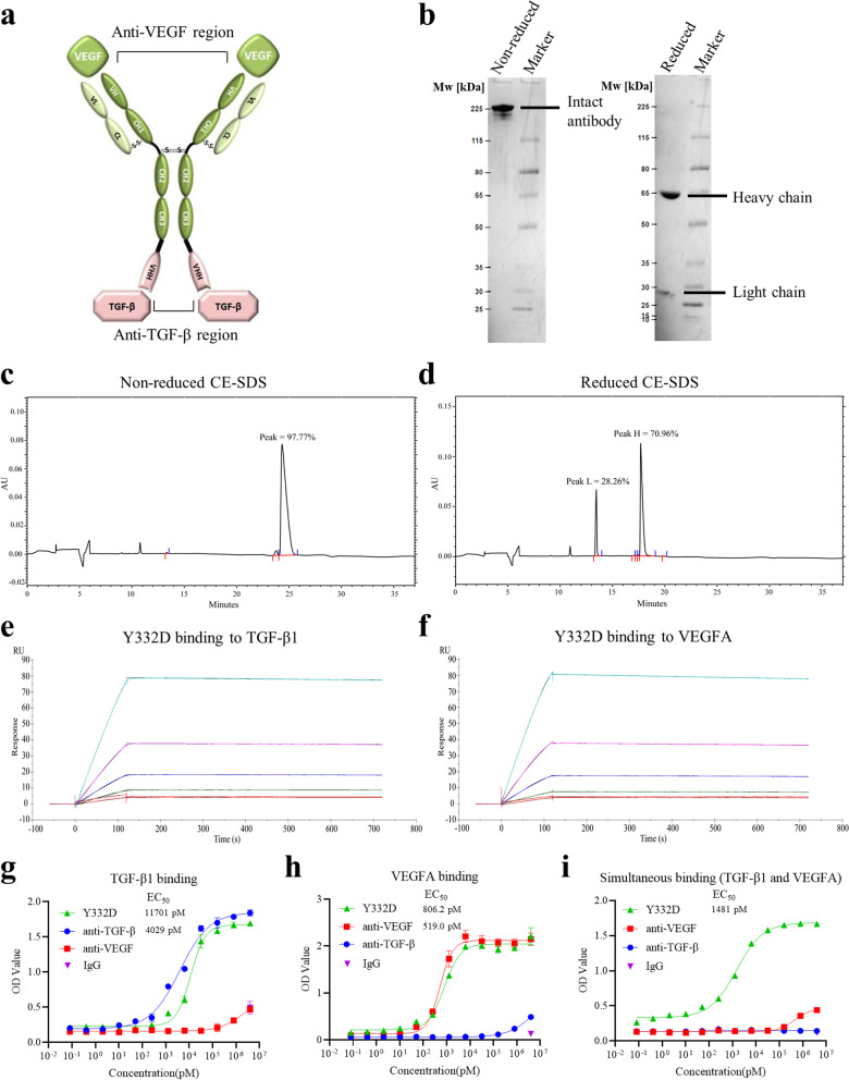
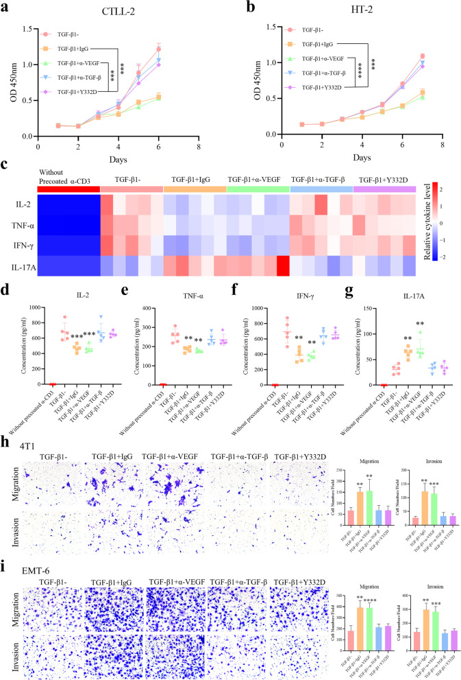
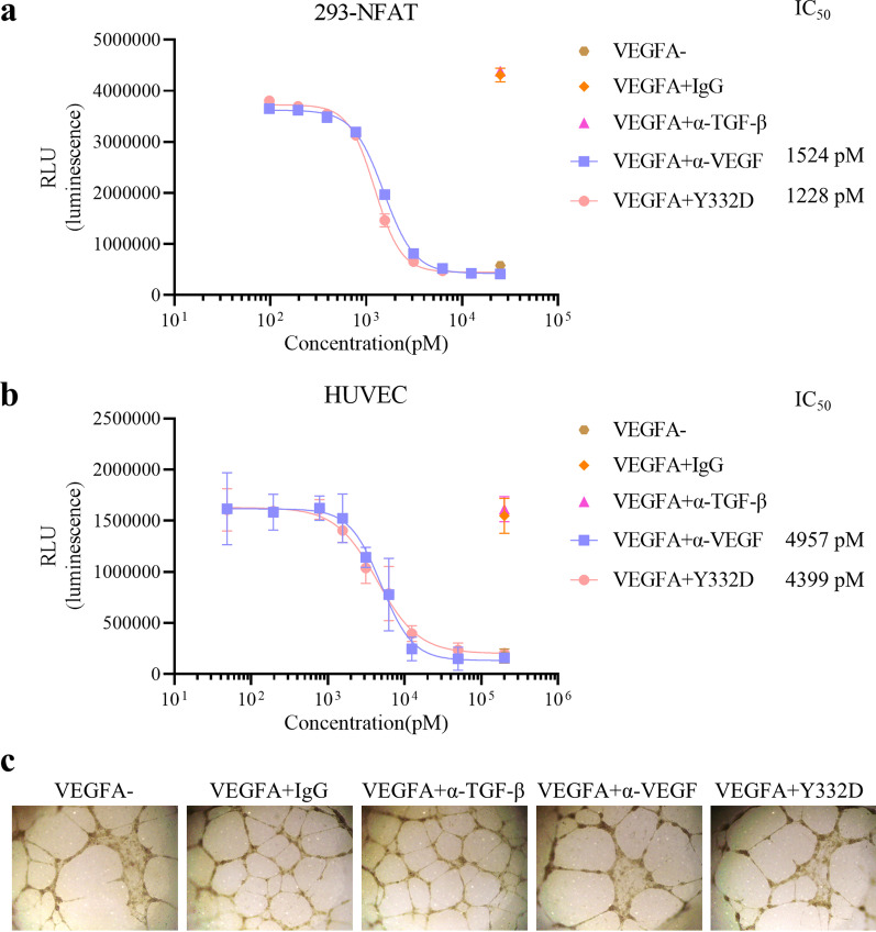
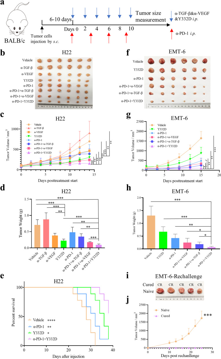
Methods: We developed a novel anti-TGF-β/VEGF bispecific antibody Y332D based on the Nano-YBODY™ technology platform. The CCK-8, flow cytometry, SBE4 luciferase reporter assay, western blotting and transwell assays were used to measure the biological activities of the anti-TGF-β moiety. The NFAT luciferase reporter assay, luminescent cell viability assay and tube formation assay were used to measure the biological activities of the anti-VEGF moiety. The in vivo anticancer efficacy of Y332D alone or in combination with PD-1 blockade was evaluated in H22, EMT-6, 4T1, and AKT/Ras-driven murine hepatocellular carcinoma tumor models. Immunofluorescent staining, flow cytometry, RNA-seq and quantitative RT-PCR were adopted to analyze the alterations in the tumor microenvironment.

Results: Y332D could maintain specific binding affinities for TGF-β and VEGFA. Y332D almost entirely counteracted the in vitro biological functions of TGF-β and VEGFA, including immunosuppression, activated TGF-β signaling, epithelial-mesenchymal transition (EMT), activated VEGF/VEGFR signaling, HUVEC proliferation and tube formation. The in vivo experiment data demonstrated that Y332D was more effective in inhibiting tumor growth and metastasis than anti-TGF-β and anti-VEGF monotherapies. In combination therapies, Y332D plus PD-1 blockade exhibited the most potent and durable anticancer effect. Mechanistically, Y332D plus PD-1 blockade upregulated the density and function of tumor-infiltrating lymphocytes and exerted reinvigorated antitumor immunity.

Conclusion: Y332D could simultaneously block TGF-β and VEGF signalings. In comparison with the monotherapies, Y332D combined with PD-1 blockade exerts superior antitumor effect through improving immune microenvironment.

Keywords: Bispecific antibody; Cancer immunotherapy; PD-1; TGF-β; The tumor microenvironment; VEGF.


Conflict of interest statement

QH, RY, LZ, JS, JZ and PZ were employees of Wuhan YZY Biopharma Co., Ltd.

Figures

Fig. 1
Fig. 1
The structure characteristics of Y332D and the binding affinity of Y332D to TGF-β and VEGF. a Schematic representation of Y332D. Y332D is designed as a tetravalent and symmetric bispecific antibody that contains two anti-VEGF regions and two anti-TGF-β regions. The long chain of Y332D consists of five domains: VHb, CH1, CH2, CH3, VHH. The short chain of Y332D contains two domains: VLb, CL. The Fc region of Y332D is an engineered hybrid fragment: the CH2 domain is from IgG2, and the CH3 domain is from IgG1. b The non-reduced and reduced SDS-PAGE analysis of Y332D. Under nonreducing conditions, a single band was displayed. Two bands were observed under reducing conditions, representing the heavy and light chains. c, d The non-reduced and reduced CE-SDS analysis of Y332D. One peak was observed in the non-reduced CE-SDS, and two peaks were detected in the reduced CE-SDS. The purity of Y332D was more than 97%. e The results of SPR assay to detect the binding kinetics of Y332D to TGF-β1. f The results of SPR assay to detect the binding kinetics of Y332D to VEGFA. g The ELISA binding affinity of serially diluted Y332D or controls to plate-coated TGF-β1. h The ELISA binding affinity of serially diluted Y332D or controls to plate-coated VEGFA. i The simultaneous binding activity of Y332D to TGF-β1 and VEGFA by ELISA. Serially diluted Y332D or controls were incubated with plate-coated TGF-β1. Then, VEGFA-Biotin and peroxidase-conjugated streptavidin were added sequentially. Bars, SDs
Fig. 2
Fig. 2
Y332D counteracted TGF-β1-induced inhibition of T cell proliferation and activation as well as epithelial-mesenchymal transition (EMT). a, b CCK-8 assays were performed to show the antagonistic effect of Y332D on TGF-β1-hampered proliferation in T cells. 1 × 103 CTLL-2 and HT-2 cells were seeded in 96-well plates. Then, 5 ng/ml TGF-β1 plus 106 pM antibodies or control were added. Cell viability was continuously monitored by CCK-8 reagent. cg Multi-cytokine assay was performed to analyze the effect of Y332D on the alteration of TGF-β1-caused cytokine secretion during T cell activation. Murine T cells were obtained from the isolation of splenocytes from C57BL/6 mice. T cells (1 × 106/ml) supplemented with anti-CD28 (3 μg/ml), TGF-β1 (5 ng/ml) and 106 pM antibodies or control were cultured in 96 well flat-bottom plates precoated with anti-CD3 (3 μg/ml). After 4 days, the cellular supernatants were harvested to measure cytokines concentration. h, i Transwell migration/invasion assays were performed to demonstrate the antagonistic effect of Y332D on TGF-β-enhanced tumor cell motility. 4T1 and EMT-6 mammary tumor cells were cultured in RPIM-1640 with 1% FBS and treated with 5 ng/ml TGF-β1 plus 106 pM antibodies or untreated for 96 h. Then, about 5 × 104 cells in 100 µl RPIM-1640 supplemented with 1%FBS were seeded in the upper chambers. The lower chambers were added with 600 µl of RPIM-1640 containing 10% FBS. After incubation for 24 h, the migratory and invasive cells were fixed with 4% paraformaldehyde and stained with 0.1% crystal violet. Bars, SDs; **p < 0.01, ***p < 0.001, and ****p < 0.0001 denote the significant difference relative to Y332D treatment. α-TGF-β: anti-TGF-β, α-VEGF: anti-VEGF
Fig. 3
Fig. 3
The antagonistic effect of Y332D on the activation of VEGF/VEGFR pathway, VEGFA-induced proliferation and tube formation in HUVECs. a NFAT-luciferase reporter assay was performed to show the blockade effect of Y332D on VEGF/VEGFR pathway. HEK-293 cells overexpressing VEGFR2 were transfected with the lentiviral vectors carrying the NFAT and luciferase gene (NFAT-RE-Luci) to construct stable transfected cell lines 293-NFAT. 293-NFAT cells were cultured in 2% FBS-DMEM with VEGFA (20 ng/ml) and serially diluted Y332D or controls for 6 h. Then, the luminescence was detected. b Luminescent cell viability assay was performed to measure the inhibitory effect of Y332D on VEGFA-induced HUVEC proliferation. 5 × 103 HUVECs were seeded in 96-well plates overnight at 37 °C. Then, the medium was replaced with endothelial cell basal medium mixed with VEGFA (50 ng/ml) and serially diluted Y332D or controls. Cell viability was detected after incubation at 37 °C for 72 h. c Tube formation assay was performed to show the inhibitory effect of Y332D on VEGFA-induced vessel-like tube formation. 2 × 104 HUVECs were seeded in 96 well flat-bottom plates after plates were precoated with 50 μl Matrigel for 30 min at 37 °C. The cells were incubated in endothelial cell complete medium mixing with 100 ng/ml VEGFA and 106 pM antibodies or control for 12 h at 37 °C. Then, HUVECs were fixed with 4% paraformaldehyde for 15 min. The images of tube-like structures were captured with inverted microscope. Bars, SDs; α-TGF-β: anti-TGF-β, α-VEGF: anti-VEGF
Fig. 4
Fig. 4
Y332D inhibited tumor growth in murine tumor models, and the combination of Y332D and PD-1 blockade demonstrated synergistic antitumor effects. Tumor volume (TV) of tumor-bearing mice was measured every other day or every two days. Mice were euthanatized when TV exceeded 2500 mm3 or the study ended. a Model establishment and treatment schedule of H22 and EMT-6 tumor models. 8.7 mg/kg α-PD-1 were administrated every two days by intraperitoneal injection for four times. Equivalent mole hIgG (8.7 mg/kg), α-VEGF (8.7 mg/kg), α-TGF-β (6 mg/kg), Y332D (10 mg/kg) were administrated on alternate days by intraperitoneal injection for six times. bd 5 × 105 H22 cells were inoculated subcutaneously in the right groin of BALB/c mice on day 0. Start of treatment on day 6. The representative tumors image, tumor growth curve, tumor weight of H22-bearing mice receiving α-PD-1 plus Y332D or controls treatment were shown. e The overall survival curves of H22-bearing mice receiving α-PD-1 plus Y332D or controls treatment were shown. fh 5 × 105 EMT-6 cells were inoculated in the right mammary fat pad of BALB/c mice on day 0. Start of treatment on day 10. The representative tumors image, tumor growth curve and tumor weight of EMT-6-bearing mice receiving α-PD-1 plus Y332D or controls treatment were shown. i, j The representative image and tumor growth curve of EMT-6 tumors in the rechallenge assay were shown. *p < 0.05, **p < 0.01, ***p < 0.001, and ****p < 0.0001 denote the significant difference relative to Y332D or Y332D plus anti-PD-1 treatment. CR: complete regression. Bars, SDs; α-PD-1: anti-PD-1, α-TGF-β: anti-TGF-β, α-VEGF: anti-VEGF
Fig. 5
Fig. 5
The combination of Y332D and PD-1 blockade demonstrated synergistic antitumor efficacy in AKT/Ras-driven murine hepatocellular carcinoma tumor model. a Model establishment and treatment schedule of AKT/Ras-driven murine hepatocellular carcinoma tumor model. The plasmid encoding myr-AKT1 and/or NRasV12 along with sleeping beauty transposase were injected into the lateral tail vein of C57BL/6 mice by hydrodynamic injection on day 0. Treatment was started on day 26 (5 mice for each group). All tumor-bearing mice were randomly divided into six groups (Vehicle, Y332D, α-PD-1, α-PD-1 plus α-VEGF, α-PD-1 plus α-TGF-β, α-PD-1 plus Y332D). 8.7 mg/kg α-PD-1 were administrated every two days by intraperitoneal injection for four times. Equivalent mole hIgG (8.7 mg/kg), α-VEGF (8.7 mg/kg), α-TGF-β (6 mg/kg), Y332D (10 mg/kg) were administrated on alternate days by intraperitoneal injection for six times. 40 days after injection, the mice were euthanized, and the liver tissues were collected. b, c The representative liver tumor images and liver weight of AKT/Ras-driven murine hepatocellular carcinoma mice receiving α-PD-1 plus Y332D or controls treatment were shown. d The overall survival curves of AKT/Ras-driven murine hepatocellular carcinoma mice receiving α-PD-1 plus Y332D or controls treatment were shown. e The representative H&E staining images of liver tissues of mice receiving the treatment of combination therapies or controls. The size of the scale bar in the immunofluorescence images refer to 5 mm or 250 μm. *p < 0.05, **p < 0.01, and ****p < 0.0001 denote the significant difference relative to Y332D plus anti-PD-1 treatment. Bars, SDs. α-PD-1: anti-PD-1, α-TGF-β: anti-TGF-β, α-VEGF: anti-VEGF
Fig. 6
Fig. 6
Immunofluorescence staining to measure the status of epithelial-mesenchymal transition (EMT) of cancer cells, carcinoma-associated fibroblast (CAF) and tumor angiogenesis in H22 tumor model. The representative images and quantitative analysis of ae EMT-related markers, including anti-E-cadherin staining, anti-Vimentin staining, anti-N-cadherin staining, f CAF marker, anti-α-SMA staining, g Anti-CD31 staining. The size of the scale bar in the immunofluorescence images refer to 100 μm. Bars, SDs; ***p < 0.001, and ****p < 0.0001 denote the significant difference relative to Y332D treatment. ns: not significant, α-TGF-β: anti-TGF-β, α-VEGF: anti-VEGF
Fig. 7
Fig. 7
Immunofluorescence staining and flow cytometry assay to analyze tumor-infiltrating lymphocytes in H22 tumor model. Mice were euthanatized when the study ended. The harvested tumor tissues were subjected to immunofluorescence and flow cytometry. a The representative immunofluorescence images and quantitative analysis of tumor-infiltrating CD8+ T cells. Harvested tumor tissues were dissociated with Collagenase B and hyaluronidase to prepare single-cell suspensions. Then, the cells were fluorescently stained with the detection antibodies. The representative images and quantitative analysis of tumor-infiltrating b, h CD8+ T cells, c, i Ki67+CD8+ T cells, d, j CD25+CD8+ T cells, e, k CD69+CD8+ T cells, f, l Granzyme B+CD8+ T cells, g, m IFN-γ+CD8+ T cells. The proportion of tumor-infiltrating lymphocytes in the total live cells was calculated. The size of the scale bar in the immunofluorescence images refer to 100 μm. Bars, SDs; *p < 0.05, **p < 0.01, ***p < 0.001, and ****p < 0.0001 denote the significant difference relative to combination treatment. ns: not significant, α-PD-1: anti-PD-1
Fig. 8
Fig. 8
RNA-seq assay to explore the immune profile of H22 tumors after different treatments. a Heat map to represent the differentially expressed genes among four groups. b Heat map to represent the expression levels of cytotoxicity-related genes (Gzma, Gzmb, Prf1, Ifng, Tnf, etc.). c Heat map to represent the expression levels of chemokines (Ccl11, Cxcl9, Cxcl11, Cxcl12, Cxcl16). d, e Heat map to represent the expression levels of signature genes in T cells and NK cells, and signature scores were calculated to quantify. f–h The top 10 significantly enriched immune-related Gene Ontology (GO) terms (α-PD-1 + Y332D vs. Vehicle; α-PD-1 + Y332D vs. α-PD-1; α-PD-1 + Y332D vs. Y332D). i, j Quantitative RT-PCR validation of selected genes identified by RNA-seq. Bars, SDs; *p < 0.05 and **p < 0.01 denote the significant difference relative to combination treatment. α-PD-1: anti-PD-1
Fig. 9
Fig. 9
Schematic diagram demonstrating the synergistic antitumor immune effect of Y332D plus PD-1 blockade. a Y332D restored the cytotoxic effects of TGF-β-suppressed CD8+ T cells and inhibited TGF-β-mediated collagen production in cancer-associated fibroblasts (CAF) cells. b Y332D promoted T cells infiltration through vascular normalization. c Y332D in combination with PD-1 blockade synergistically enhanced antitumor immunity

References

    1. Boussiotis VA. Molecular and biochemical aspects of the PD-1 checkpoint pathway. N Engl J Med. 2016;375:1767–1778. doi: 10.1056/NEJMra1514296. - DOI - PMC - PubMed
    1. Liu Z, Yu X, Xu L, Li Y, Zeng C. Current insight into the regulation of PD-L1 in cancer. Exp Hematol Oncol. 2022;11:44. doi: 10.1186/s40164-022-00297-8. - DOI - PMC - PubMed
    1. Yang X, Ma L, Zhang X, Huang L, Wei J. Targeting PD-1/PD-L1 pathway in myelodysplastic syndromes and acute myeloid leukemia. Exp Hematol Oncol. 2022;11:11. doi: 10.1186/s40164-022-00263-4. - DOI - PMC - PubMed
    1. Freeman GJ, Long AJ, Iwai Y, Bourque K, Chernova T, Nishimura H, et al. Engagement of the PD-1 immunoinhibitory receptor by a novel B7 family member leads to negative regulation of lymphocyte activation. J Exp Med. 2000;192:1027–1034. doi: 10.1084/jem.192.7.1027. - DOI - PMC - PubMed
    1. Niu M, Liu Y, Yi M, Jiao D, Wu K. Biological characteristics and clinical significance of soluble PD-1/PD-L1 and exosomal PD-L1 in cancer. Front Immunol. 2022;13:827921. doi: 10.3389/fimmu.2022.827921. - DOI - PMC - PubMed

Publication types

Substances