. 2023 Aug 13;18(1):100.
doi: 10.1186/s13020-023-00806-9.

Bojungikgi-tang improves skin barrier function and immune response in atopic dermatitis mice fed a low aryl hydrocarbon receptor ligand diet

Affiliations

Bojungikgi-tang improves skin barrier function and immune response in atopic dermatitis mice fed a low aryl hydrocarbon receptor ligand diet

You Mee Ahn et al. Chin Med. .

Abstract

Background: The aryl hydrocarbon receptor (AhR) is a transcription factor that plays a crucial role in regulating the immune system and maintaining skin barrier function. AhR signaling is pivotal in the pathogenesis of inflammatory diseases such as atopic dermatitis (AD), and the absence of AhR ligands further contributes to the progression or worsening of AD symptoms.

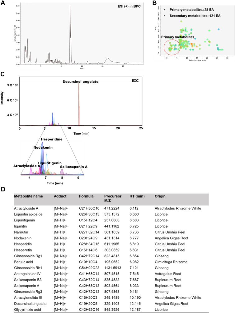
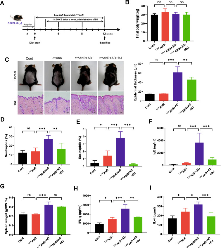
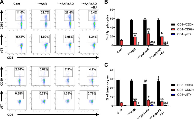
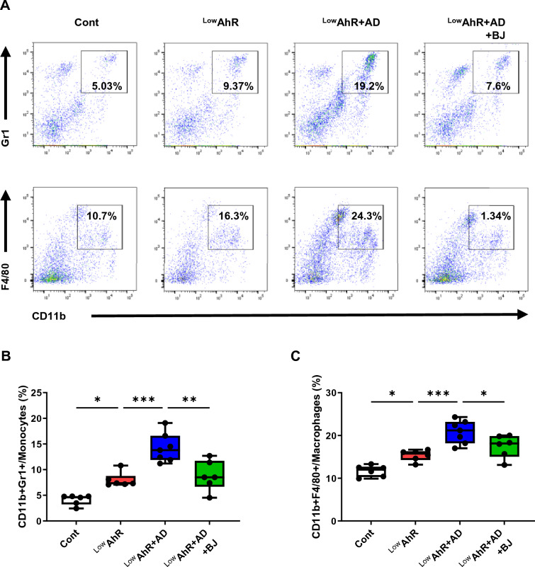
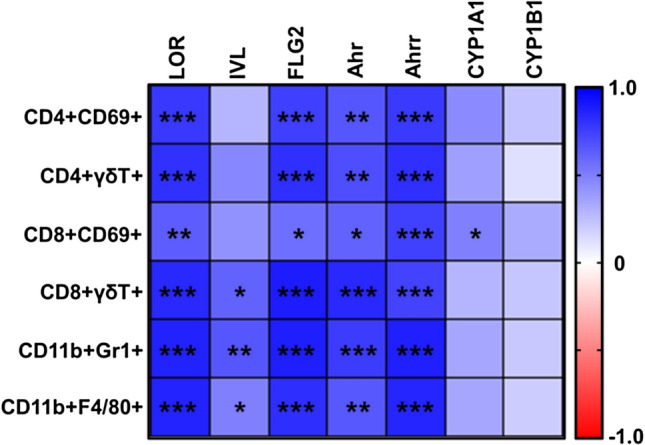
Methods: AD was induced with 2,4-dinitrochlorobenzene (DNCB), and Bojungikgi-tang (BJIKT) was administered orally daily for 10 weeks. Serum IgE, splenocyte IL-4, and IFN-γ levels, skin barrier genes, and AhR target gene expressions were analyzed using RNA-sequencing analysis. Spleen tissues were extracted for fluorescence-activated cell sorting (FACS) analysis to analyze the effect of BJIKT on immune responses. A correlation analysis was conducted to analyze the correlation between immune markers and skin barrier genes and AhR target genes.

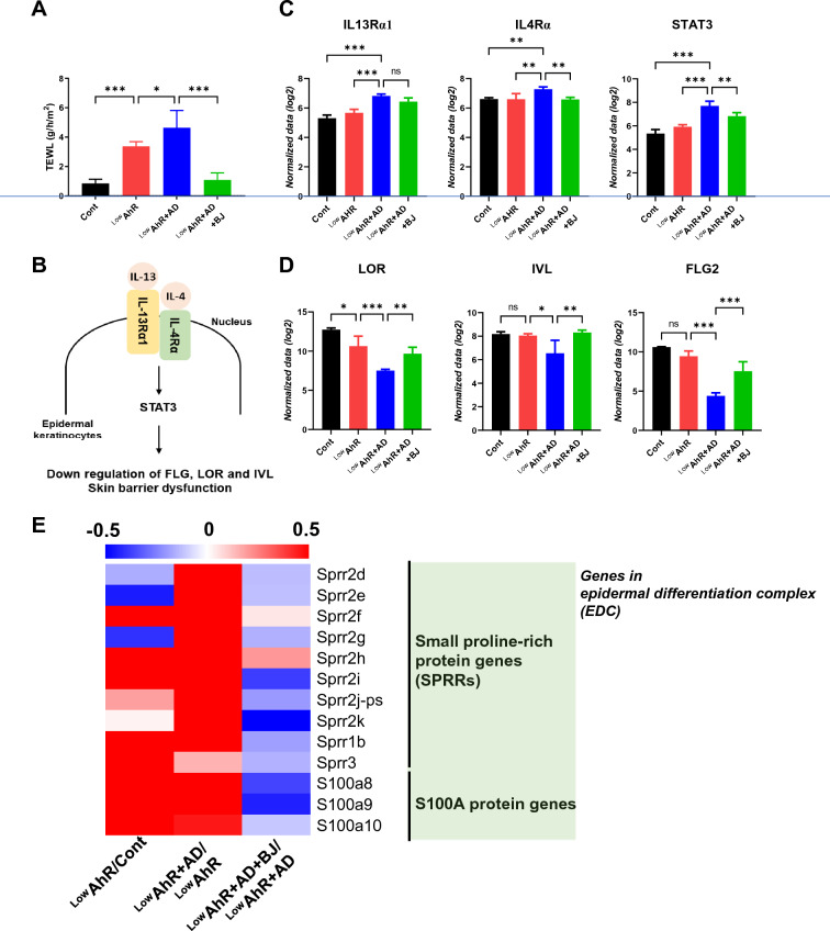
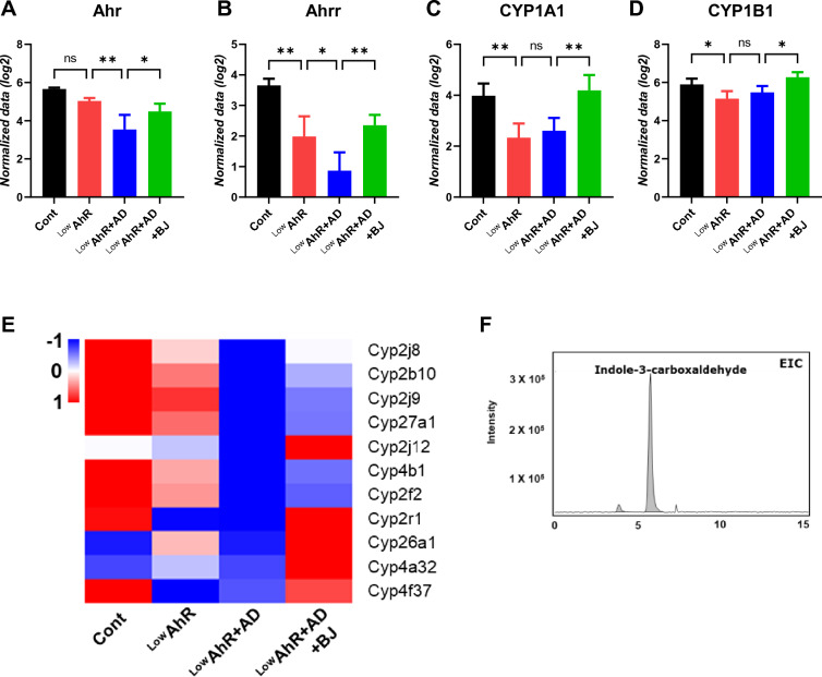
Results: BJIKT effectively improved AD symptoms in AD mice fed a low AhR ligand diet by reducing neutrophil and eosinophil counts, lowering IgE levels in the blood, and decreasing IL-4 and IFN-γ levels in the splenocytes. Additionally, BJIKT significantly reduced epithelial skin thickness and transepidermal water loss (TEWL) values and reversed the decreased expression of skin barrier genes. BJIKT also considerably altered the expression of AhR target genes, including Ahr, Ahrr, cytochrome P450 1A1 (CYP1A1), and CYP1B1. Furthermore, AhR target pathway genes were negatively correlated with immune cell subtypes, including CD4 + and CD8 + T cells and macrophages (CD11b + F4/80 +) at the systemic level.

Conclusions: BJIKT can regulate AhR activation and may help reduce inflammation in AD by regulating the expression of skin barrier genes and immune responses.

Keywords: Atopic dermatitis; BJIKT; Immune response; Inflammation; Low-AhR ligand diet; Skin barrier.


Conflict of interest statement

The authors declare no competing interests.

Figures

Fig. 1
Fig. 1
Chemical profiling of Bojungikgi-tang (BJIKT). A Base peak chromatograms (BPC); B A peak spot graph representing identified metabolites; C Extracted ion chromatograms (EIC) of six major constituents in BJIKT; D List of major chemical constituents in BJIKT
Fig. 2
Fig. 2
Effects of BJIKT on 2,4-dinitrochlorobenzene (DNCB)-induced atopic dermatitis (AD) symptoms in AD mice fed a low AhR ligand diet. A Timeline for AD mice fed a low AhR ligand diet and treatment with BJIKT; B Final body weight was scored 10 weeks after AD induction; C Evaluation of the total dorsal skin and epithelial thickness by histological analysis; measurement of the levels of D neutrophils and (E) eosinophils by CBC analysis; F IgE plasma levels; G spleen weight; and the levels of H interferon-gamma (IFN)-γ and I interleukin (IL)-4 in splenocytes. Number of experiments conducted for group comparisons: Cont, n = 6; LowAhR, n = 6; LowAhR + AD, n = 7; and LowAhR + AD + BJ, n = 6. The values are expressed as the means ± standard error of the mean (SEM). *p < 0.05, **p < 0.01, ***p < 0.001 according to a one-way ANOVA followed by Tukey’s multiple comparisons
Fig. 3
Fig. 3
Effects of BJIKT on skin barrier dysfunction in AD mice fed a low AhR ligand diet. A Measurement of transepidermal water loss (TEWL) was performed at the end of the experiments after sensitization using TM300; RNA-seq analysis of the expression levels of the differentially expressed genes B Activation of the IL‐13/4‐STAT3 axis inhibits the expression of FLG and LOR (downregulation of barrier); C IL4Rα, IL13Rα1, STAT3 in the skin; D loricrin (LOR), involucrin (IVL), and filaggrin (FLG2) in the skin; E A representative heatmap of the RNA-seq analysis of skin tissues. Number of experiments conducted for group comparisons: Cont, n = 4; LowAhR, n = 4; LowAhR + AD, n = 4; and LowAhR + AD + BJ, n = 4. The values are expressed as the means ± SEM. *p < 0.05, **p < 0.01, ***p < 0.001 according to a one-way ANOVA followed by Tukey’s multiple comparisons
Fig. 4
Fig. 4
Upregulation of AhR target genes by BJIKT in AD mice fed a low AhR ligand diet. RNA-seq analysis of the relative expression of AhR target genes (A) Ahr, (B) Ahrr, (C) CYP1A1, and (D) CYP1B1 in the skin. (E) A representative heatmap of the differentially expressed Cytochrome P450 family genes in the skin. F TheeExtracted ion chromatograms (EIC) of indole-3-carboxaldehyde as an AhR ligand in BJIKT-treated cells. Number of experiments conducted for group comparisons: Cont, n = 4; LowAhR, n = 4; LowAhR + AD, n = 4; and LowAhR + AD + BJ, n = 4. The values are expressed as mean ± SEM. *p < 0.05, **p < 0.01, according to a one-way ANOVA followed by Tukey’s multiple comparisons
Fig. 5
Fig. 5
The effects of BJIKT on CD4 + and CD8 + T cell populations in AD mice fed a low AhR ligand diet. Fluorescence-activated cell sorting of CD4 and CD8 T cell populations in the spleen. A Gating strategy for identification of CD4 + /CD3 + , CD4 + /CD69 + , CD4 + /γδT + , CD8 + /CD3 + , CD8 + /CD69 + , and CD8 + /γδT + in splenocytes; percentage of B CD4 + T cells (CD4 + /CD3 + , CD4 + /CD69 + , CD4 + /γδT +) and C CD8 + T cells (CD8 + /CD3 + , CD8 + /CD69 + , CD8 + /γδT +) in splenocytes. Number of experiments conducted for group comparisons: Cont, n = 6; LowAhR, n = 6; LowAhR + AD, n = 7; and LowAhR + AD + BJ, n = 6. The values are expressed as the means ± SEM. *p < 0.05, **p < 0.01 vs Cont; #p < 0.05, ##p < 0.01, ###p < 0.001 vs. LowAhR; $p < 0.05, $$p < 0.01, $$$p < 0.001 vs. LowAhR + AD. Only these two groups were compared and the significance was marked
Fig. 6
Fig. 6
The effects of BJIKT on myeloid-derived suppressor cells (MDSCs) and macrophage cells in AD mice fed a low AhR ligand diet. Fluorescence-activated cell sorting of MDSCs (CD11b + Gr1 +) and macrophage (CD11b + F4/80 +) populations in the spleen. A Density plots showing gating strategies for MDSCs (CD11b + Gr1 +) and macrophage (CD11b + F4/80 +) cells; they show differences in percentages of B CD11b + Gr1 + and C CD11b + F4/80 + cells. Number of experiments conducted for group comparisons: Cont, n = 6; LowAhR, n = 6; LowAhR + AD, n = 7; and LowAhR + AD + BJ, n = 6. The values are expressed as the means ± SEM. *p < 0.05, **p < 0.01, ***p < 0.001, according to a one-way ANOVA followed by Tukey’s multiple comparisons
Fig. 7
Fig. 7
Correlation analysis between alterations in immune markers and skin barrier and AhR target gene expression levels after BJIKT administration. The heatmap shows the correlations between immune markers and barrier genes (LOR, IVL, and FLG2) and AhR target genes (Ahr, Ahrr, CYP1A1, and CYP1B1) levels in the skin. Pearson’s r values were determined using GraphPad Prism 9. The red color indicates a negative correlation. Correlation is significant at the *p < 0.05, **p < 0.01, ***p < 0.001 level

References

    1. Esser C, Rannug A. The aryl hydrocarbon receptor in barrier organ physiology, immunology, and toxicology. Pharmacol Rev. 2015;67:259–279. doi: 10.1124/pr.114.009001. - DOI - PubMed
    1. Paternoster L, Savenije OEM, Heron J, Evans DM, Vonk JM, Brunekreef B, et al. Identification of atopic dermatitis subgroups in children from 2 longitudinal birth cohorts. J Allergy Clin Immunol. 2018;141:964–971. doi: 10.1016/j.jaci.2017.09.044. - DOI - PMC - PubMed
    1. Van Den Bogaard EH, Podolsky MA, Smits JP, Cui X, John C, Gowda K, et al. Genetic and pharmacological analysis identifies a physiological role for the AHR in epidermal differentiation. J Invest Dermatol. 2015;135:1320–1328. doi: 10.1038/jid.2015.6. - DOI - PMC - PubMed
    1. Furue M, Tsuji G, Mitoma C, Nakahara T, Chiba T, Morino-Koga S, et al. Gene regulation of filaggrin and other skin barrier proteins via aryl hydrocarbon receptor. J Dermatol Sci. 2015;80:83–88. doi: 10.1016/j.jdermsci.2015.07.011. - DOI - PubMed
    1. Fritsche E, Schäfer C, Calles C, Bernsmann T, Bernshausen T, Wurm M, et al. Lightening up the UV response by identification of the arylhydrocarbon receptor as a cytoplasmatic target for ultraviolet B radiation. Proc Natl Acad Sci USA. 2007;104:8851–8856. doi: 10.1073/pnas.0701764104. - DOI - PMC - PubMed