. 2023 Aug 14;8(1):299.
doi: 10.1038/s41392-023-01558-6.

High-density lipoprotein regulates angiogenesis by long non-coding RNA HDRACA

Affiliations

High-density lipoprotein regulates angiogenesis by long non-coding RNA HDRACA

Zhi-Wei Mo et al. Signal Transduct Target Ther. .

Abstract

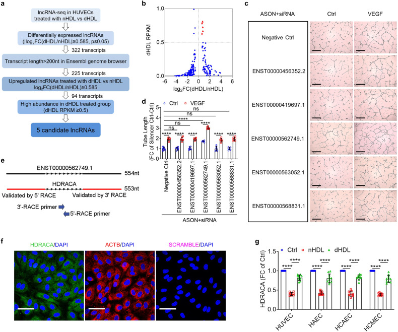
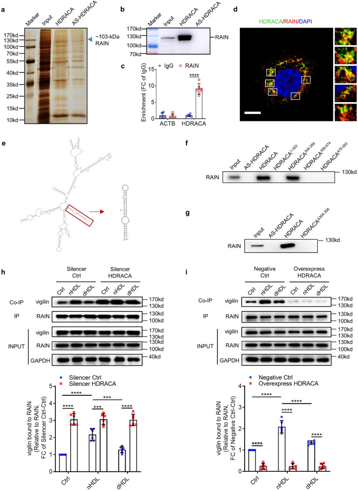
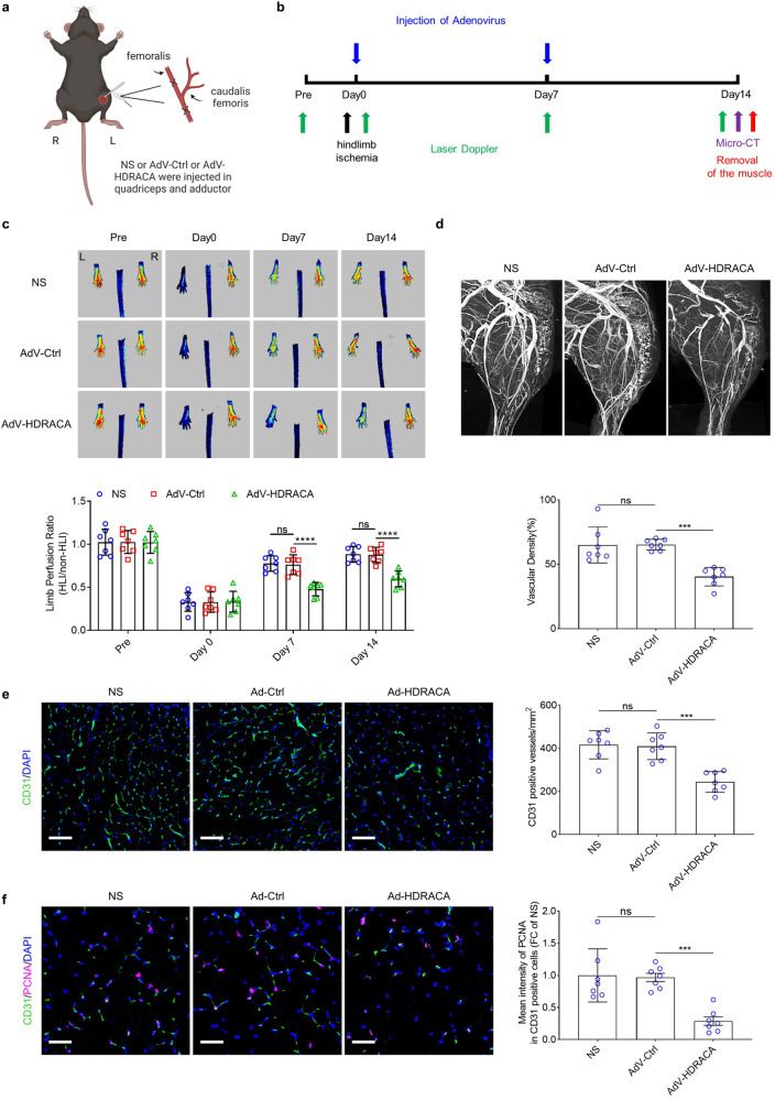
Normal high-density lipoprotein (nHDL) can induce angiogenesis in healthy individuals. However, HDL from patients with coronary artery disease undergoes various modifications, becomes dysfunctional (dHDL), and loses its ability to promote angiogenesis. Here, we identified a long non-coding RNA, HDRACA, that is involved in the regulation of angiogenesis by HDL. In this study, we showed that nHDL downregulates the expression of HDRACA in endothelial cells by activating WW domain-containing E3 ubiquitin protein ligase 2, which catalyzes the ubiquitination and subsequent degradation of its transcription factor, Kruppel-like factor 5, via sphingosine 1-phosphate (S1P) receptor 1. In contrast, dHDL with lower levels of S1P than nHDL were much less effective in decreasing the expression of HDRACA. HDRACA was able to bind to Ras-interacting protein 1 (RAIN) to hinder the interaction between RAIN and vigilin, which led to an increase in the binding between the vigilin protein and proliferating cell nuclear antigen (PCNA) mRNA, resulting in a decrease in the expression of PCNA and inhibition of angiogenesis. The expression of human HDRACA in a hindlimb ischemia mouse model inhibited the recovery of angiogenesis. Taken together, these findings suggest that HDRACA is involved in the HDL regulation of angiogenesis, which nHDL inhibits the expression of HDRACA to induce angiogenesis, and that dHDL is much less effective in inhibiting HDRACA expression, which provides an explanation for the decreased ability of dHDL to stimulate angiogenesis.


Conflict of interest statement

The authors declare no competing interests.

Figures

Fig. 1
Fig. 1
nHDL and dHDL cause different expression of HDRACA in endothelial cells. a Selection strategy of lncRNA in HUVECs treated with nHDL or dHDL. b Scatter plot of differentially expressed lncRNAs in HUVECs induced by nHDL and dHDL (Transcripts length > 200 nt in Ensembl genome browser database). Red symbols represent the 5 candidate lncRNAs. c, d Tube formation assays showed the effects of respectively knocking down 5 candidate lncRNAs using specific ASONs (66 nM) and siRNAs (33 nM) on angiogenesis. The representative images (c) and quantification (d) are shown. Scale bars, 500 μm. e RACE assays measured the full length of ENST00000562749.1 termed HDRACA. f FISH assays showed the subcelluar location of HDRACA in HUVECs. Representative images of HDRACA (green), ACTB (red) and Scramble-ISH (magenta) are shown. ACTB probes are shown as a positive control, and Scramble-ISH probes are shown as a negative control. The nuclei were stained with DAPI (blue). Scale bars, 50 μm. g RT-qPCR confirmed the expression of HDRACA in multiple kinds of endothelial cells treated with nHDL or dHDL. Data are presented as the mean ± SD. For (c) and (d), n = 6. For (g), n = 10. ****p < 0.0001; ns not significant
Fig. 2
Fig. 2
HDL-bound S1P regulates the transcription of HDRACA in the endothelial cells by affecting the ubiquitination of KLF5. a RT-qPCR showed the effect of W146 on HDRACA levels in HUVECs treated with nHDL or dHDL. b The S1P content in nHDL (n = 20) and dHDL (n = 20). c The levels of HDRACA in HUVECs treated with increasing concentrations of S1P (from 0.5 μM to 8 μM) were assayed by RT-qPCR. d The levels of HDRACA in HUVECs treated with S1P (1 μM), r-ApoM (1 μM), and r-ApoM-S1P (1 μM) were assayed by RT-qPCR. e The middle tracks display the online dataset of KLF5 ChIP-seq in GM12878 (blue; GSE127670), H3K4me3 ChIP-seq in HUVECs (red; GSM945181, GSM733673, GSE96250) and H3K27ac ChIP-seq in HUVECs (yellow; GSM733691). The purple arrow indicates the putative KLF5 binding site in HDRACA promoter based on the conserved KLF5 binding motif (shown on the top) and peaks. f The mRNA levels of KLF5 in HUVECs treated with nHDL or dHDL were assayed by RT-qPCR. g Immunoblot of KLF5 in HUVECs treated with nHDL or dHDL. The representative plots (left) and quantification (right) are shown. h KLF5 ubiquitination in HUVECs treated with nHDL or dHDL was assayed by immunoprecipitation (IP) and immunoblotting (IB). MG132 was added to inhibit KLF5 degradation. i RT-qPCR confirmed the mRNA levels of HDRACA in HUVECs transfected with negative control siRNA or KLF5-siRNAs. j ChIP-RT-qPCR analysis (right) for KLF5 binding to the HDRACA promoter in HUVECs. Normalized data are shown as percentages of the input controls. KLF5 binding to BECN1 promoter served as a positive control. The whole-cell lysate (INPUT) and immunoprecipitated proteins (IP) of each immunoprecipitation were analyzed by immunoblotting (left) for KLF5 or IgG. k Luciferase reporter assays for HUVECs with endogenous KLF5 expression transfected with pGL4.1 reporter plasmids containing deletion HDRACA promoter constructs. HUVECs transfected with a blank pGL4.1 plasmid served as a negative control. l Luciferase reporter assays for HUVECs with endogenous KLF5 expression transfected with pGL4.1 reporter plasmids containing wild type or mutated HDRACA promoters. HUVECs transfected with a blank pGL4.1 plasmid served as a negative control. Data are presented as the mean ± SD. For all the experiments, n = 6. **p < 0.01; ***p < 0.001; ****p < 0.0001; ns not significant
Fig. 3
Fig. 3
nHDL and dHDL differently regulate HDRACA expression to affect endothelial cell proliferation and tube formation. a Volcano plot shows differentially expressed genes in HUVECs after treatment with lncRNA Smart Silencer HDRACA (Silencer HDRACA) compared with lncRNA smart silencer negative control (Silencer Ctrl), n = 3. b, c Flow cytometric analysis of HUVECs showing the cell cycle at 30 min after addition of PI/RNAse and 48 h after lncRNA Smart Silencer transfection (b) or 72 h after lentivirus vector transfection (c). The representative plots (left and middle) and quantification (right) are shown. d, e The representative images (left) and quantification (right) of EdU incorporation assay in HUVECs treated with VEGF or nHDL or dHDL after silencing (d) or overexpressing (e) HDRACA. The proliferative HUVECs were labeled with EdU (red) and the nuclei were stained with Hoechst 33342 (blue). Scale bars, 100 μm. f, g The representative images (left) and quantification (right) of tube formation assay in HUVECs treated with VEGF or nHDL or dHDL after silencing (f) or overexpressing (g) HDRACA. Scale bars, 500 μm. Data are presented as the mean ± SD. For (bg), n = 6. *p < 0.05; **p < 0.01; ***p < 0.001; ****p < 0.0001
Fig. 4
Fig. 4
HDRACA hinders the interaction between RAIN and vigilin in endothelial cells. a Silver staining of proteins bound to HDRACA or antisense for HDRACA in HUVECs after RNA pull-down. RAIN (arrow) was identified by mass spectrometry. AS-HDRACA means antisense for HDRACA. b Immunoblotting for RAIN on protein lysate from HUVECs after RNA pull-down. c RIP assays demonstrated the interaction of HDRACA and RAIN in HUVECs. ACTB was used as a negative control. d Confocal images showing the colocalization of HDRACA (green) and RAIN (red) in HUVECs. The nuclei were stained with DAPI (blue). Scale bars, 10 μm. e Predicted secondary structure of HDRACA and HDRACA304–358 by Mfold software. f RNA pull-down assays determined the interaction between HDRACA domain constructs and RAIN. g RNA pull-down analysis of biotinylated HDRACA nucleotides 304–358 deletion construct and RAIN. h, i Immunoprecipitation analysis of the interaction of RAIN and vigilin in HUVECs treated with nHDL or dHDL after silencing (h) or overexpressing (i) HDRACA. Representative plots (up) and quantitation (down) are shown. Data are presented as the mean ± SD. For (bi), n = 6. ***p < 0.001; ****p < 0.0001
Fig. 5
Fig. 5
HDRACA increases the inhibitory effect of vigilin on PCNA expression in endothelial cells. a Heatmap shows the differentially expressed cell cycle genes after transfection with Silencer Ctrl or Silencer HDRACA. b Cell cycle genes expression in HUVECs after transfection with negative control siRNA or HDRACA-siRNAs were determined by RT-qPCR. c RT-qPCR determined the expression of cell cycle genes upregulated>1.5 Fold in panel (b) after transfection with negative control siRNA or RAIN-siRNAs. d The expression of cell cycle genes differentially expressed in panel (c) after transfection with negative control siRNA or Vigilin-siRNAs was determined by RT-qPCR. Data in panel (bd) were normalized to the negative control siRNA group (siCtrl), and the dotted lines represent the value of the siCtrl. e The binding of vigilin protein with PCNA, TFDP1, YWHAH, PKMYT1, or CDC6 mRNAs in HUVECs was determined by RIP assays. f, g The representative plots (left) and quantitation (right) of immunoblot analysis of PCNA in HUVECs treated with nHDL or dHDL after silencing (f) or overexpressing (g) HDRACA. h The representative images (left) and quantification (right) of EdU incorporation assay in HUVECs treated with VEGF or nHDL or dHDL after transfection with negative control siRNA or PCNA-siRNA (siPCNA-2). The proliferative HUVECs were labeled with EdU (red) and the nuclei were stained with Hoechst 33342 (blue). Scale bars, 100 μm. i The representative images (left) and quantification (right) of tube formation assay in HUVECs treated with VEGF or nHDL or dHDL after transfection with negative control siRNA or PCNA-siRNA (siPCNA-2). Scale bars, 500 μm. Data are presented as the mean ± SD. For (bi), n = 6. For (bd), *p < 0.05 compared to siCtrl. For (ei), **p < 0.01; ***p < 0.001; ****p < 0.0001
Fig. 6
Fig. 6
Ectopic expression of human HDRACA in mouse endothelial cells inhibits angiogenesis. a RT-PCR indicated the presence of human HDRACA in mFAECs transfected with lentiviruses. b The representative images (up) and quantification (down) of EdU incorporation assays in mFAECs treated with or without VEGF after overexpressing HDRACA. The proliferative mFAECs were labeled with EdU (red) and the nuclei were stained with Hoechst 33342 (blue). Scale bars, 100 μm. c The representative images (up) and quantification (down) of tube formation assays in mFAECs treated with or without VEGF after overexpressing HDRACA. Scale bars, 500 μm. d Immunoblotting for RAIN on protein lysate from mFAECs after RNA pull-down with biotin-labeled HDRACA or HDRACA antisense (AS-HDRACA). e RIP assays demonstrated the interaction between HDRACA and RAIN in mFAECs transfected with HDRACA-overexpressing lentiviruses. Gapdh was used as a negative control. Data are presented as the mean ± SD. n = 6. ****p < 0.0001
Fig. 7
Fig. 7
HDRACA inhibits angiogenesis in vivo. a Schematic graphic of the ischemia model used in this study. b Overview of experimental process in vivo. c Representative images (up) and quantification (down) of laser doppler flow before and at various time points after femoral artery ligation in mice treated with normal saline (NS), control adenovirus vector (AdV-Ctrl), or adenovirus vector carrying HDRACA (AdV-HDRACA). R (right) and L (left) foot. d Representative images (up) and quantification (down) of micro-CT analysis at day 14 after femoral artery ligation in mice treated with NS or AdV-Ctrl or AdV-HDRACA. e Representative images (left) and quantification (right) of CD31 staining (green) at day 14 after femoral artery ligation in mice treated with NS, AdV-Ctrl or AdV-HDRACA. The nuclei were stained with DAPI (blue). Scale bars, 100 μm. f Representative images (left) and quantification (right) of CD31 (green) and PCNA (magenta) co-staining at day 14 after femoral artery ligation in mice treated with NS, AdV-Ctrl or AdV-HDRACA. The nuclei were stained with DAPI (blue). Scale bars, 50 μm. Data are presented as the mean ± SD. For (cf), n = 7. ***p < 0.001; ****p < 0.0001; ns not significant
Fig. 8
Fig. 8
HDRACA is upregulated in lower extremity artery intima of patients with ASO. a Representative images of co-stain for CD31 (green) with KLF5 (red; up), HDRACA (red; middle) and PCNA (red; down) in artery tissues from donors for organ transplantation (Ctrl; n = 10) and patients with ASO (n = 10). The nuclei were stained with DAPI (blue). Scale bars, 50 μm. b Quantification of KLF5, HDRACA, or PCNA mean fluorescence intensity in CD31 positive cells of artery tissues from Ctrl and ASO groups. c Correlation between S1P content in HDL and HDRACA mean fluorescence intensity in CD31 positive cells of artery tissues. n = 20. d Graphical illustration of HDL-HDRACA regulatory mechanism. (1) nHDL-bound S1P interacts with S1P1, leading to the phosphorylation and activation of WWP2. The activated WWP2 catalyzes the ubiquitination and subsequent degradation of KLF5, resulting in reduced KLF5-induced HDRACA transcription. Consequently, non-HDRACA-bound RAIN increases and interacts with vigilin to inhibit its binding to PCNA mRNA, leading to increased stability of PCNA mRNA, increased PCNA expression, and increased PCNA-induced endothelial cells proliferation and tube formation, ultimately promoting angiogenesis. (2) dHDL does not inhibit KLF5-induced HDRACA transcription via S1P1-WWP2 signaling pathway, due to the loss of S1P. HDRACA binds to RAIN, which disrupts the interaction between RAIN and vigilin, resulting in the binding of vigilin and PCNA mRNA, thereby decreasing the stability of PCNA mRNA and PCNA expression. As a result, dHDL fails to promote angiogenesis by increasing the expression of PCNA and PCNA-induced proliferation and tube formation. Data are presented as the mean ± SD. ***p < 0.001; ****p < 0.0001; ns not significant

References

    1. Toyota E, et al. Vascular endothelial growth factor is required for coronary collateral growth in the rat. Circulation. 2005;112:2108–2113. - PubMed
    1. Sabra M, et al. Clinical application of novel therapies for coronary angiogenesis: overview, challenges, and prospects. Int. J. Mol. Sci. 2021;22:3722. - PMC - PubMed
    1. Kontush A, Lhomme M, Chapman MJ. Unraveling the complexities of the HDL lipidome. J. Lipid Res. 2013;54:2950–2963. - PMC - PubMed
    1. Rohatgi A, Westerterp M, von Eckardstein A, Remaley A, Rye KA. HDL in the 21st century: a multifunctional roadmap for future HDL research. Circulation. 2021;143:2293–2309. - PMC - PubMed
    1. Li HM, et al. Angiogenic and antiangiogenic mechanisms of high density lipoprotein from healthy subjects and coronary artery diseases patients. Redox Biol. 2020;36:101642. - PMC - PubMed

Publication types

Substances