Review
. 2023 Jun 9;5(9):100811.
doi: 10.1016/j.jhepr.2023.100811. eCollection 2023 Sep.

Non-alcoholic fatty liver disease and diabetes mellitus as growing aetiologies of hepatocellular carcinoma

Affiliations
Review

Non-alcoholic fatty liver disease and diabetes mellitus as growing aetiologies of hepatocellular carcinoma

Stephanie Talamantes et al. JHEP Rep. .

Abstract

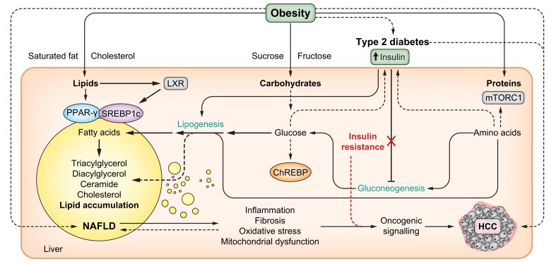
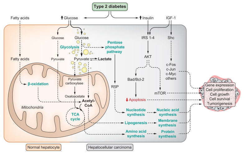
Obesity-related complications such as non-alcoholic fatty liver disease (NAFLD) and type 2 diabetes (T2D) are well-established risk factors for the development of hepatocellular carcinoma (HCC). This review provides insights into the molecular mechanisms that underlie the role of steatosis, hyperinsulinemia and hepatic inflammation in HCC development and progression. We focus on recent findings linking intracellular pathways and transcription factors that can trigger the reprogramming of hepatic cells. In addition, we highlight the role of enzymes in dysregulated metabolic activity and consequent dysfunctional signalling. Finally, we discuss the potential uses and challenges of novel therapeutic strategies to prevent and treat NAFLD/T2D-associated HCC.

Keywords: Hepatocellular carcinoma; hepatocyte transformation; non-alcoholic fatty liver disease; obesity; type 2 diabetes.


Figures

Fig. 1
Fig. 1
Obesity and overnutrition expose the liver to an overload of energetic fuels (lipids, carbohydrates and proteins) that can affect hepatic energy metabolism with pathological consequences. Several transcription factors are known to act as sensors of nutrients, such as PPAR-γ, which is activated by lipophilic ligands such as PUFAs; SREBP1c, which is activated by LXR in response to insulin, PUFAs and oxysterols; ChREBP, which is activated by glucose-6-phosphate; or mTORC1, which is activated in response to amino acids. These and other transcription factors regulate the expression of enzymes and signalling proteins required to execute and coordinate major metabolic pathways. The consequence of chronic aberrant activation of these transcription factors, associated with hyperinsulinemia during T2D predispose the liver to steatosis, inflammation, fibrosis, oxidative stress and mitochondrial dysfunction. These factors facilitate oncogenic transformation and HCC development. Selective insulin resistance confers liver resistance to the inhibitory action of insulin on gluconeogenesis, while the sensitivity of the liver to the stimulatory effect of insulin over lipogenesis remains. Different types of nutrients provided by the diet can accelerate metabolic dysfunction, including nutrients that are abundant in industrialised highly palatable and caloric foods such as saturated fat, cholesterol, sucrose and fructose. ChREB, carbohydrate-responsive element-binding protein; HCC, hepatocellular carcinoma; LXR, liver X receptor; mTORC1, mechanistic target of rapamycin complex 1; NAFLD, non-alcoholic fatty liver disease; PUFA, polyunsaturated fatty acid; T2D, type 2 diabetes; PPAR-γ, peroxisome proliferator-activated receptor gamma; SREBP1c, sterol regulatory element-binding protein 1c.
Fig. 2
Fig. 2
NAFLD and T2D are characterised by increased hepatic inflammation and oxidative stress, contributing to HCC development. The population of resident and recruited immune cells in the liver is dynamic during the progression of NASH. Different cell types can participate in the inflammatory response. Pro-inflammatory cytokines such as IL-6 and TNF-α, are released by different tissue sources and drive an inflammatory response, including oncogenic STAT3 activation. An excess of dietary fats and simple carbohydrates favour body and hepatic fat accumulation and alters specific immune cell populations in the liver. Tumour suppressive CD4+ T cells are depleted by nutrient overload and ROS-dependent mechanisms. Hepatic resident KCs are also depleted in NAFLD and replaced by two subsets of pro-inflammatory recruited macrophages: monocyte-derived KCs and hepatic lipid-associated macrophages. These macrophages co-localize with fibrotic liver regions with activated hepatic stellate cells. In association with hepatic steatosis and inflammation, continuously high ROS levels lead to oxidative stress and liver damage that is strongly associated with HCC development. CD4, cluster of differentiation 4; HCC, hepatocellular carcinoma; IL-6, interleukin-6; KCs, Kupffer cells; NAFLD, non-alcoholic fatty liver disease; ROS, reactive oxygen species; STAT-3, signal transducer and activator of transcription 3; TNF-α, tumor necrosis factor alpha; T2D, type 2 diabetes.
Fig. 3
Fig. 3
Hepatocyte transformation in NAFLD/T2D. Normal hepatocytes prefer β-oxidation of fatty acids as a source of energy, as well as relatively low glucose uptake and oxidation. This is especially true during fasting conditions and this preference is influenced by hepatic zonation. Transformed hepatocytes exhibit high glucose uptake and rely on aerobic glycolysis as a source of energy. Pyruvate is preferentially converted into lactate, instead of being oxidised in the mitochondria. High fat intake rewires hepatocyte energy metabolism to favour glucose uptake and its utilisation as a source of energy through aerobic glycolysis and lactate production. Glucose can be used as a carbon source for biosynthetic reactions in rapidly growing tissues, as well as in cell signalling and maintenance of the redox state. High glucose uptake also sustains the substrate requirement for the pentose phosphate pathway. This is important for ribulose-5-phosphate synthesis, which is required for nucleotide biosynthesis and nucleic acid replication. Pyruvate carboxylase is induced by high-fat intake, favouring the entrance of pyruvate into the TCA cycle as oxaloacetate to maintain anaplerotic reactions required for amino acid biosynthesis. Acetyl-CoA is converted into fatty acids through lipogenesis. The pool of intracellular fatty acids (from lipogenesis and extrahepatic tissues) is used for phospholipid biosynthesis to build biological membranes. Hyperinsulinemia associated with T2D, in combination with the action of the hepatokine IGF-1, can lead to dysfunctional signalling pathways involved in cell survival, apoptosis, and stress responses. Together, these cellular and metabolic changes can represent advantages for cancer cells, allowing them to sustain rapid growth and proliferation. Bad, Bcl-2-associated agonist of cell death; Bcl-2, B-cell lymphoma 2; HCC, hepatocellular carcinoma; IGF-1, insulin-like growth factor 1; IRS1-4, insulin receptor substrate 1-4; mTOR, mammalian target of rapamycin; NAFLD, non-alcoholic fatty liver disease; Shc, Src homology 2 domain-containing transforming protein; T2D, type 2 diabetes; TCA, tricarboxylic acid.

References

    1. Sung H., Ferlay J., Siegel R.L., Laversanne M., Soerjomataram I., Jemal A., et al. Global cancer statistics 2020: GLOBOCAN estimates of incidence and mortality worldwide for 36 cancers in 185 countries. CA Cancer J Clin. 2021;71:209–249. - PubMed
    1. Llovet J.M., Kelley R.K., Villanueva A., Singal A.G., Pikarsky E., Roayaie S., et al. Hepatocellular carcinoma. Nat Rev Dis Primers. 2021;7:6. - PubMed
    1. Sanyal A.J., Harrison S.A., Ratziu V., Abdelmalek M.F., Diehl A.M., Caldwell S., et al. The natural history of advanced fibrosis due to nonalcoholic steatohepatitis: data from the simtuzumab trials. Hepatology. 2019;70:1913–1927. - PubMed
    1. Rinella M.E., Neuschwander-Tetri B.A., Siddiqui M.S., Abdelmalek M.F., Caldwell S., Barb D., et al. AASLD Practice Guidance on the clinical assessment and management of nonalcoholic fatty liver disease. Hepatology. 2023;77:1797–1835. - PMC - PubMed
    1. Huang D.Q., Singal A.G., Kono Y., Tan D.J.H., El-Serag H.B., Loomba R. Changing global epidemiology of liver cancer from 2010 to 2019: NASH is the fastest growing cause of liver cancer. Cell Metab. 2022;34:969–977 e962. - PMC - PubMed

LinkOut - more resources