. 2023 Jul 24;9(8):e18599.
doi: 10.1016/j.heliyon.2023.e18599. eCollection 2023 Aug.

Combination of human platelet lysate and 3D gelatin scaffolds to enhance osteogenic differentiation of human amniotic fluid derived mesenchymal stem cells

Affiliations

Combination of human platelet lysate and 3D gelatin scaffolds to enhance osteogenic differentiation of human amniotic fluid derived mesenchymal stem cells

Kantirat Yaja et al. Heliyon. .

Abstract

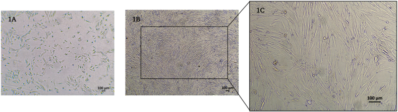
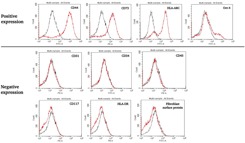
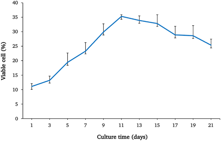
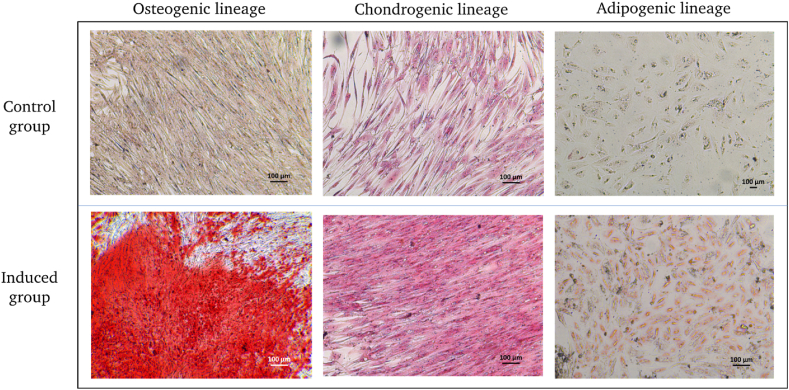
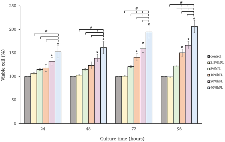
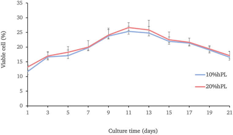
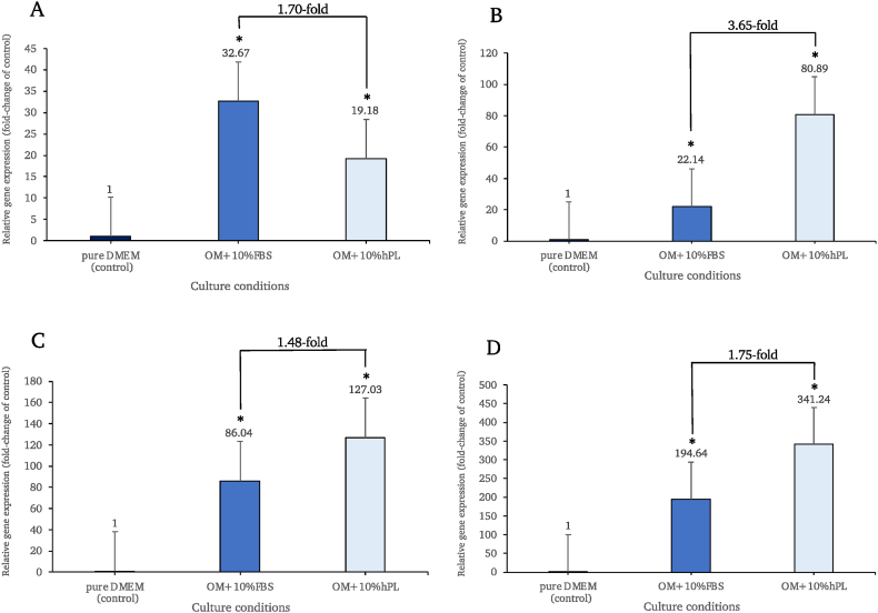
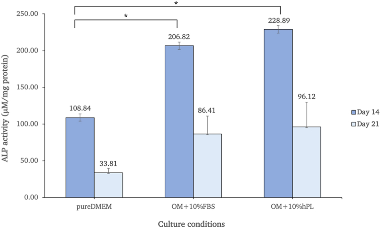
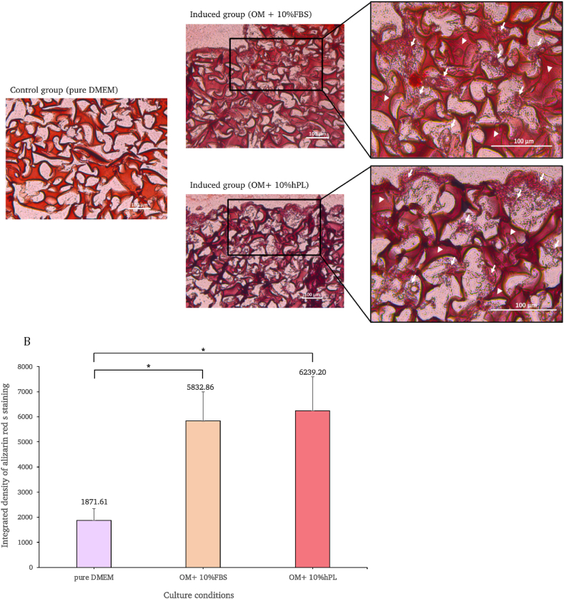
Bone disorders are major health issues requiring specialized care; however, the traditional bone grafting method had several limitations. Thus, bone tissue engineering has become a potential alternative. In therapeutic treatments, using fetal bovine serum (FBS) as a culture supplement may result in the risk of contamination and host immunological response; therefore, human platelet lysate (hPL) has been considered a viable alternative source. This study attempted to compare the effectiveness and safety of different culture supplements, either FBS or hPL, on the osteoblastic differentiation potential of mesenchymal stem cells derived from human amniotic fluid (hAF-MSCs) under a three-dimensional gelatin scaffold. The results indicate that hAF-MSCs have the potential to be used in clinical applications as they meet the criteria for mesenchymal stem cells based on their morphology, the expression of a particular surface antigen, their proliferation ability, and their capacity for multipotent differentiation. After evaluation by MTT and Alamar blue proliferation assay, 10% of hPL was selected. The osteogenic differentiation of hAF-MSCs under three-dimensional gelatin scaffold using osteogenic-induced media supplemented with hPL was achievable and markedly stimulated osteoblast differentiation. Moreover, the expressions of osteoblastogenic related genes, including OCN, ALP, and COL1A1, exhibited the highest degree of expression under hPL-supplemented circumstances when compared with the control and the FBS-supplemented group. The induced cells under hPL-supplemented conditions also presented the highest ALP activity level and the greatest degree of calcium accumulation. These outcomes would indicate that hPL is a suitable substitute for animal derived serum. Importantly, osteogenic differentiation of human amniotic fluid derived mesenchymal stem cells using hPL-supplemented media and three-dimensional scaffolds may open the door to developing an alternative construct for repairing bone defects.

Keywords: Human amniotic fluid mesenchymal stem cells; Human platelet lysate; Osteoblast-like cells; Osteogenic differentiation; Scaffold; Tissue engineering.


Conflict of interest statement

The authors declare that they have no known competing financial interests or personal relationships that could have appeared to influence the work reported in this paper.

Figures

Fig. 1
Fig. 1
Morphology of hAF cells at the 1st passage (A) and hAF cells at the 2nd passage (B) with 4× magnification (1B) and 10× magnification (1C).
Fig. 2
Fig. 2
Flow cytometry histogram of the expressions of the cell surface protein markers of hAF cells during the 2nd passage. The hAF cells exhibited a positive degree of expression of CD44, CD73, HLA-ABC, and Oct-4. On the other hand, the cells were found to be negative for CD31, CD34, CD45, CD117, HLA-DR, and fibroblast surface protein markers.
Fig. 3
Fig. 3
The growth curve and viability of the hAF cells over 21 days. Data are presented as mean ± SEM values.
Fig. 4
Fig. 4
Accordingly, hAF differentiation potential assay after 21 days of specific induction.
Fig. 5
Fig. 5
Viability of hAF-MSCs exposed to hPL (2.5–40%) in DMEM for 24, 48,72, and 96 h. The cells cultured in pure DMEM were used as the control. Data are presented as mean ± SEM values. * Denoted for statistical significance when compared with the control group (p < 0.05) # Denoted for statistical significance among the groups (p < 0.05).
Fig. 6
Fig. 6
The growth curve and viability of the hAF cells cultured in media supplemented with either 10% hPL or 20% hPL oveer 21 days. Data are presented as mean ± SEM values.
Fig. 7
Fig. 7
Expression levels of osteoblastogenic related genes, including RUNX2 (A), OCN (B), ALP (C), and COL1A1 (D), were normalized to GAPDH and were relative to the control group on day 14. * Denoted for statistical significance when compared with the control group (p < 0.05).
Fig. 8
Fig. 8
ALP activity of hAF-MSCs cultivated for 14 and 21 days under different culture medium conditions.
Fig. 9
Fig. 9
Calcium deposition of the control group and the osteogenic-induced group under scaffold culture conditions was identified with alizarin red S staining. Arrows are used to represent deposited calcium, while arrowheads are used to represent the scaffold (A). In the graph, the staining intensities of the cells cultured with pure DMEM, and the osteogenic-induced medium supplemented with 10% FBS and 10% hPL under scaffold culture conditions, are compared. One-way ANOVA was used to identify differences between the groups and the conditions (p < 0.05) (B). (For interpretation of the references to color in this figure legend, the reader is referred to the Web version of this article.)

References

    1. Dimitriou R., Jones E., McGonagle D., Giannoudis P.V. Bone regeneration: current concepts and future directions. BMC Med. 2011;9:66. - PMC - PubMed
    1. Mistry A.S., Mikos A.G. Tissue engineering strategies for bone regeneration. Adv. Biochem. Eng. Biotechnol. 2005;94:1–22. - PubMed
    1. Waese E.Y., Kandel R.A., Stanford W.L. Application of stem cells in bone repair. Skeletal Radiol. 2008;37:601–608. - PubMed
    1. Khaled E.G., Saleh M., Hindocha S., Griffin M., Khan W.S. Tissue engineering for bone production- stem cells, gene therapy and scaffolds. Open Orthop. J. 2011;5(Suppl 2):289–295. - PMC - PubMed
    1. Maxson S., Lopez E.A., Yoo D., Danilkovitch-Miagkova A., Leroux M.A. Concise review: role of mesenchymal stem cells in wound repair. Stem Cells Transl Med. 2012;1:142–149. - PMC - PubMed